

IMEMR
EMRO
Volume 7, Issue 2 (2026)
3 2026, 7(2): 101-106 |
Back to browse issues page
Request Type:




History
Received: 2026/03/10 | Accepted: 2026/06/9 | Published: 2026/06/21
Received: 2026/03/10 | Accepted: 2026/06/9 | Published: 2026/06/21
How to cite this article
Naser H, Al-Semaiil F, Mohammed M, Abbas B. Isolation and Identification of Proteus mirabilis from Clinical Samples Using the VITEK-2 Automated System. 3 2026; 7 (2) :101-106
URL: http://jccs.daneshafarand.org/article-8-87254-en.html
URL: http://jccs.daneshafarand.org/article-8-87254-en.html
Download citation:
BibTeX | RIS | EndNote | Medlars | ProCite | Reference Manager | RefWorks
Send citation to:


Rights and permissions
BibTeX | RIS | EndNote | Medlars | ProCite | Reference Manager | RefWorks
Send citation to:


1- Department of Pathological Analyses, Faculty of Science, Al-Kunooze University, Basra, Iraq
2- Department of Science, College of Education Open (Al-Dujail Study Branch), Al-Dujail, Iraq
3- Department of Pathological Analysis, Faculty of Health and Medical Techniques, Al-Kunooze University, Basra, Iraq, Department of Pathological Analysis, Faculty of Health and Medical Techniques, Al-Kunooze University, Street/Al-Zaytoon, Basra, Iraq. Postal Code: 610001 (basilabbas63@gmail.com)
Keywords:
| Abstract (HTML) (90 Views)
Full-Text: (6 Views)
Introduction
Proteus mirabilis is a Gram-negative bacterium belonging to the family Enterobacteriaceae and is recognized as an important opportunistic pathogen in humans. It is particularly associated with urinary tract infections (UTIs), although it can also be isolated from wound infections and other clinical conditions. Its clinical importance is related to a combination of virulence-associated properties, including motility, adhesion, biofilm formation, and urease activity, which contribute to its ability to colonize and persist in the urinary tract [1-3].
A characteristic feature of P. mirabilis is its pronounced swarming motility on solid media, which facilitates surface colonization and contributes to the spread of bacterial populations. The organism can also form biofilms and express several virulence determinants that support its persistence under different environmental conditions. Recent studies have demonstrated associations among biofilm formation, swarming behavior, virulence factors, and antimicrobial resistance, emphasizing the biological versatility of this species [4-6].
The identification of P. mirabilis in clinical microbiology commonly involves a combination of colony characteristics, microscopic examination, and biochemical reactions. Important phenotypic characteristics include glucose fermentation, hydrogen sulfide production, urease activity, and decarboxylase reactions. In particular, urease production is closely related to the pathogenic behavior of P. mirabilis because urease hydrolyzes urea and contributes to an increase in local pH, thereby favoring the formation of urinary stones and persistence within the urinary tract [2, 7]. Recent clinical studies have continued to use conventional biochemical testing in combination with automated identification systems to identify P. mirabilis isolates [8, 9].
Although conventional biochemical tests remain useful, interpreting multiple reactions can be laborious, particularly when a large number of clinical isolates must be processed. Variations in individual biochemical reactions may also occur among isolates of the same bacterial species, making identification based on a single characteristic less reliable. Recent investigations of P. mirabilis have demonstrated phenotypic and genetic diversity among clinical isolates, including differences in virulence-associated characteristics and antimicrobial resistance profiles [10-12]. Therefore, the combined interpretation of several biochemical reactions can provide a more informative phenotypic profile for bacterial identification.
Automated identification systems have been increasingly incorporated into clinical microbiology laboratories to facilitate standardized and rapid bacterial identification. The VITEK 2 system uses predefined biochemical reaction patterns to compare a tested isolate with a database of bacterial profiles and provides an identification along with a probability or confidence value. Recent clinical studies have used VITEK 2 in combination with conventional culture and biochemical characteristics to identify P. mirabilis, including investigations conducted in Iraq and other clinical settings [8, 9, 13]. The continued use of automated identification in recent clinical studies supports its practical relevance for the routine characterization of Gram-negative bacterial isolates.
The VITEK 2 GN card is designed to identify clinically important Gram-negative organisms using a panel of biochemical reactions. The resulting pattern can be represented as a numerical biotype or bionumber, while the identification probability indicates how closely the observed biochemical profile corresponds to the profiles contained in the system database. Consequently, evaluating the individual biochemical reactions underlying an identification may provide additional information beyond the final species name alone. Recent applications of automated identification systems have emphasized the importance of combining instrument-generated identifications with phenotypic and clinical information when characterizing bacterial isolates [9, 13, 14].
For P. mirabilis, biochemical characteristics, such as hydrogen sulfide production, urease activity, glucose fermentation, and decarboxylase reactions are particularly relevant to phenotypic differentiation. However, the expression of individual reactions may not be completely uniform among isolates. Recent studies have reported variation in phenotypic characteristics and virulence-associated traits among P. mirabilis populations, supporting the need to examine isolate-level differences rather than assume an identical biochemical pattern for all members of the species [13, 15, 16]. Characterizing these variations may therefore improve our understanding of the biochemical profiles generated during automated identification.
Despite the widespread use of automated identification systems, relatively limited attention has been given to the detailed biochemical profiles generated by the VITEK 2 GN card for individual clinical P. mirabilis isolates, particularly regarding the consistency of individual reactions and variation in their bionumbers. Recent studies have focused mainly on antimicrobial resistance, virulence determinants, epidemiology, or molecular characteristics of P. mirabilis rather than describing the complete phenotypic reaction profile associated with automated identification. Therefore, examining the biochemical reactions and identification outputs generated by VITEK 2 may provide useful phenotypic information about clinical P. mirabilis isolates [8, 11, 17, 18].
This study aimed to characterize the phenotypic identification profiles of clinical P. mirabilis isolates using the VITEK 2 GN card, with particular emphasis on identification probability, bionumbers, analysis time, and the consistency and variability of the biochemical reactions detected by the system.
Materials and Methods
This descriptive observational study was conducted in 2025 in a clinical microbiology laboratory. Over 150 urine samples were processed, and 23 consecutive Gram-negative clinical isolates recovered from routine specimens in the clinical microbiology laboratory were included in the study. The isolates were obtained using standard media, including 5% sheep blood agar, MacConkey agar, and cysteine-lactose-electrolyte-deficient (CLED) agar. The media were incubated at 35±1°C for 18-24 hours, and the isolates were processed consecutively without selection by specimen type. All isolates produced pure cultures and exhibited oxidase-negative, Gram-negative rod morphology on these media. Single colonies were subcultured to ensure purity before VITEK 2 testing.
Identification was performed using the VITEK 2 Compact instrument (bioMérieux) and the GN identification card, in accordance with the manufacturer’s instructions. A bacterial suspension equivalent to a 0.5-0.63 McFarland standard was prepared in 0.45% saline from an 18-24-h pure culture. The GN card was inoculated, sealed, and loaded into the instrument’s incubator-reader carousel. The instrument monitored each of the 47 wells fluorometrically or colorimetrically every 15 min for up to 18 hours, applied a kinetic algorithm to compare the binary profile with its internal database, and returned a final taxon identification with an associated identification probability (%), a unique 15-digit bionumber, and a total analysis time (in hours).
For each of the 23 isolates, the following parameters were manually extracted from the VITEK 2 printout: selected organism, identification probability, bionumber, total analysis time, and the binary result (+, –, or a bracketed intermediate result, such as “(+)” or “(–)”) for each of the 47 biochemical wells on the GN card. For each taxon identified with a probability of ≥90%, the results were aggregated at the species level. For P. mirabilis specifically, positivity rates for each reaction were calculated as the proportion of the nine P. mirabilis isolates that returned a positive (unbracketed “+”) result. Reactions bracketed by the instrument were considered borderline and excluded from the “definite positive” tallies. Individual bionumbers were examined for shared patterns to assess intraspecies diversity.
The aggregated VITEK 2 biochemical profile of P. mirabilis was qualitatively compared with classical descriptions of the species in the peer-reviewed literature [3].
Findings
The VITEK 2 system returned a species- or genus-level identification with a probability of ≥90% for 18 of 23 isolates (78.3%) and returned an “Unidentified Organism” status for 5 isolates (21.7%). All identifications were final: no isolate required repeat testing for technical reasons, and no instrument-reported contamination was detected. Of the 18 identified isolates, 14 (60.9% of the total cohort) were identified with a probability of ≥99%, whereas 4 (17.4%) were identified with a probability of 90–98%.
Among the 18 successfully identified isolates, the following taxa were detected: P. mirabilis (n = 9; 39.1% of the total cohort and 50.0% of identified isolates); Aeromonas sobria (n = 3; 13.0% and 16.7%, respectively); Aeromonas hydrophila/punctata (caviae) (n=1; 4.3% and 5.6%, respectively); Enterobacter cloacae complex (n=1; 4.3% and 5.6%, respectively); Plesiomonas shigelloides (n=1; 4.3% and 5.6%, respectively); Rhizobium radiobacter (n=1; 4.3% and 5.6%, respectively); Pandoraea spp. (n=1; 4.3% and 5.6%, respectively); and Bordetella bronchiseptica (n=1; 4.3% and 5.6%, respectively). The remaining five isolates (21.7% of the total cohort) were reported as unidentified. P. mirabilis was the most frequently identified taxon. All nine P. mirabilis isolates were reported with high identification probabilities: eight of nine (88.9%) at 99% probability and one (11.1%) at 96% probability. Analysis times ranged from 3.35 to 4.82 hours, with a mean of 3.84 hours. The two longest analysis times, 4.82 and 3.87 hours, were associated with additional well resolution because of minor delays in individual fluorometric endpoints; these delays were not associated with reduced identification confidence
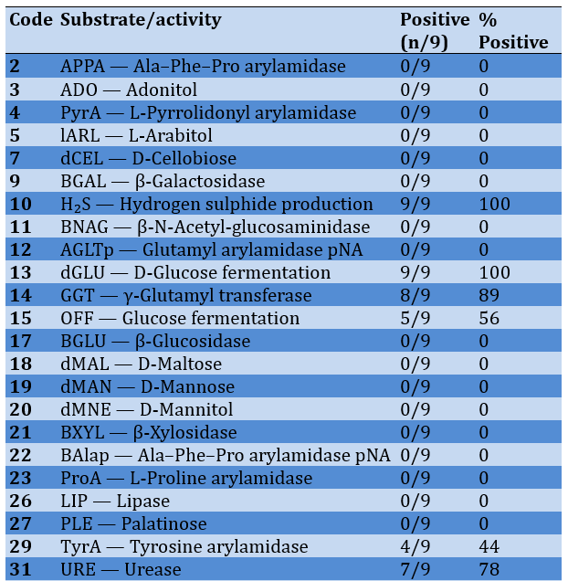
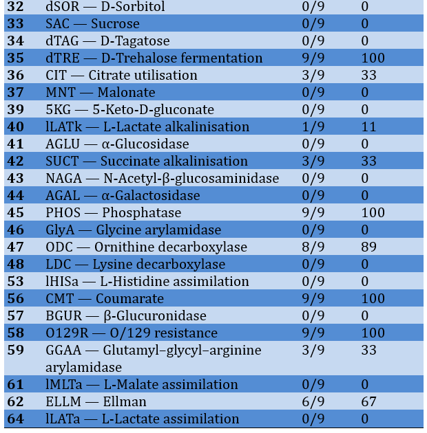
The aggregated reaction profile of the nine VITEK 2-identified P. mirabilis w:as char:acterized by uniformly positive reactions for hydrogen sulfide production (H₂S, 9/9, 100%), D-glucose fermentation (dGLU, 9/9, 100%), D-trehalose fermentation (dTRE, 9/9, 100%), phosphatase (PHOS, 9/9, 100%), the coumarate reaction (CMT, 9/9, 100%), and resistance to the vibriostatic agent O/129 (O129R, 9/9, 100%). These reactions formed the core biochemical pattern supporting assignment to P. mirabilis. All isolates were negative for the lactose-associated reactions dCEL, BGAL, and SAC. Urease was positive in 7 of 9 isolates (78%), while ornithine decarboxylase was positive in 8 of 9 (89%) and lysine decarboxylase was negative in all isolates. Thus, the predominant ODC-positive/LDC-negative pattern was consistent with the classical biochemical profile of P. mirabilis, although one isolate was ODC-negative
Most reactions were invariant across the nine isolates. In addition to the uniformly positive reactions described above, LDC was negative in all isolates. The principal variable reactions were GGT, which was positive in 8 of 9 isolates (89%); ELLM, positive in 6 of 9 (67%); OFF, positive in 5 of 9 (56%); TyrA, positive in 4 of 9 (44%); and URE, SUCT, and GGAA, each positive in 3 of 9 isolates (33%). These variations accounted for the differences observed among the P. mirabilis bionumbers but did not prevent species-level identification (Table 1).
Table 1. Consolidated biochemical profile of Proteus mirabilis isolates (n=9) on the VITEK 2 GN card
In addition to the nine P. mirabilis identifications, the VITEK 2 system returned acceptable identifications for Aeromonas sobria (n=3; identification probabilities of 95%, 95%, and 93%), Aeromonas hydrophila/punctata (caviae) (n=1; 99%), Enterobacter cloacae complex (n=1; 90%), Plesiomonas shigelloides (n=1; 99%), Rhizobium radiobacter (n=1; 91%), Pandoraea spp. (n = 1; 94%), and Bordetella bronchiseptica (n=1; 99%). The mean analysis time for this combined group, excluding P. mirabilis, was 5.18 hours, compared with 3.84 hours for the P. mirabilis isolates. The biochemical profiles and bionumbers of these isolates were consistent with the expected profiles for their respective taxa on the GN card.
Discussion
This study aimed to characterize the phenotypic identification profiles of clinical P. mirabilis isolates using the VITEK 2 GN card. The VITEK 2 GN card identified P. mirabilis with high confidence, achieving a probability of ≥96% for every P. mirabilis identification and a probability of 99% for 8 of 9 isolates (88.9%). This high level of confidence is consistent with previously published evaluations of VITEK 2 for Proteus identification, in which the species is reported to be one of the best-resolved taxa in the GN database [10, 18, 19]. The mean analysis time of 3.84 h was comparable to the rapid-identification range reported for Proteus on VITEK 2 in routine clinical use.
A noteworthy observation in the dataset was the relationship between analysis time and identification confidence. The “Unidentified Organism” group required a mean incubation time of 8.28 hours—more than twice the time required for P. mirabilis—yet still failed to match any taxon in the database. This finding suggests that the kinetic algorithm allows atypical phenotypes the maximum possible readout time before declaring the result inconclusive, reflecting the internal conservatism built into the instrument. The five unidentified bionumbers likely represent atypical biogroups that are not yet present in the VITEK 2 database and support the use of combined phenotypic–molecular workflows in which unidentified VITEK 2 isolates are resolved by 16S rRNA gene sequencing [11, 19].
The VITEK 2 biochemical profile of P. mirabilis closely conformed to the classical phenotypic description of the species. The clustered reactions with 100% consistency (H₂S⁺, D-glucose⁺, D-trehalose⁺, phosphatase⁺, CMT⁺, and O/129-R⁺), together with the 89% consistency of the ornithine decarboxylase (ODC⁺) reaction and the uniformly negative lysine decarboxylase (LDC⁻) reaction, closely corresponded to the published biochemical description of P. mirabilis: swarming, urease-positive, lactose-negative, hydrogen sulfide-producing, indole-negative, and ornithine decarboxylase-positive [19].
The differential value of the ornithine decarboxylase reaction was particularly evident in this dataset. Eight of nine P. mirabilis isolates (89%) were ODC-positive, and all nine were LDC-negative, producing the ODC⁺/LDC⁻ combination classically regarded as the single most useful biochemical discriminator between P. mirabilis and P. vulgaris, which displays the LDC⁺/ODC⁻ combination. The single ODC-negative isolate (bionumber 0017000240040211) emphasizes that P. mirabilis identification cannot rely on ODC positivity alone. However, the uniform LDC negativity across all nine isolates, together with the consistent H₂S-, D-glucose-, D-trehalose-, phosphatase-, and O/129-R-positive pattern, maintained species-level differentiation. H₂S production was uniformly positive (100%), in agreement with the established phenotype of P. mirabilis [19].
The universal absence of indole-related reactions was consistent with the classically indole-negative character of the species.
The observed variability in some biochemical reactions within the P. mirabilis cohort was itself informative. Urease activity was positive in 78% (7/9) of the isolates, with two isolates (bionumbers 0013000040042211 and 0013000040042231) lacking urease—an unusual finding because urease is typically considered a hallmark of the species [3]. Ornithine decarboxylase was positive in 89% (8/9) of the isolates, with a single isolate (bionumber 0017000240040211) lacking this reaction. This finding is consistent with the 5-15% phenotypic variability in ODC activity reported in surveys of clinical P. mirabilis collections [15, 19, 20]. γ-Glutamyl transferase showed variability (89% positivity), glucose fermentation, as detected by OFF, was variably present (56%), and tyrosine arylamidase was variably present (44%). Citrate (CIT, 33%), GGAA (33%), and ELLM (67%) showed considerable heterogeneity, and the lLATk and SUCT reactions were occasionally positive (11% and 33%, respectively). This variability is consistent with the published literature describing intraspecies biochemical variation among clinical P. mirabilis isolates [10, 15, 20] and reinforces the value of using a probabilistic, multitest database for species identification rather than relying on any single reaction. The nine distinct bionumbers generated for the nine isolates make this heterogeneity explicit at the binary-profile level.
This study has several limitations. First, the cohort size (n=23) was modest. Although the cohort included a diversity of clinically relevant Gram-negative taxa, the findings for P. mirabilis and the “Unidentified Organism” group specifically require confirmation in larger series. Second, identification of P. mirabilis in this study was based solely on VITEK 2 GN-card biochemistry, without molecular confirmation (e.g., 16S rRNA gene sequencing or rpoB sequencing) of either the P. mirabilis identifications or the “Unidentified Organism” calls. Third, because the present cohort included isolates collected during a defined period and from a single laboratory, the generalizability of the bionumber distributions and unidentified-profile patterns to other settings cannot be assured. Fourth, this study did not address potential antimicrobial susceptibility patterns among the P. mirabilis isolates, which have been reported elsewhere to be evolving toward MDR and XDR [10, 11, 19, 20]. Future studies should combine VITEK 2 identification with MALDI-TOF mass spectrometry, 16S rRNA gene sequencing, and antimicrobial susceptibility testing to provide a comprehensive characterization of clinical P. mirabilis isolates.
Conclusion
The VITEK 2 GN card provides high-confidence identification of Proteus mirabilis and demonstrates a consistent biochemical profile across the isolates.
Acknowledgments: The authors acknowledge Biovet Lab (Basrah) for providing laboratory facilities.
Ethical Permissions: The study was conducted in accordance with the Declaration of Helsinki and approved by the Ethics Committee of the University of Basrah Review Board (Protocol Code: UOB-ETH-2025-16; December 2025).
Conflicts of Interests: Authors declare there is no conflict of interest.
Authors' Contribution: Naser HA (First Author), Introduction Writer/Methodologist/Main Researcher (25%); Al-semaiil FB (Second Author), Methodologist/Assistant Researcher (25%); Mohammed MD (Third Author), Assistant Researcher/Discussion Writer/Statistical Analyst (25%); Abbas BA (Fourth Author), Introduction Writer/Methodologist/Assistant Researcher (25%)
Funding/Support: The researcher received no funding from any organization; the study was self-funded.
Proteus mirabilis is a Gram-negative bacterium belonging to the family Enterobacteriaceae and is recognized as an important opportunistic pathogen in humans. It is particularly associated with urinary tract infections (UTIs), although it can also be isolated from wound infections and other clinical conditions. Its clinical importance is related to a combination of virulence-associated properties, including motility, adhesion, biofilm formation, and urease activity, which contribute to its ability to colonize and persist in the urinary tract [1-3].
A characteristic feature of P. mirabilis is its pronounced swarming motility on solid media, which facilitates surface colonization and contributes to the spread of bacterial populations. The organism can also form biofilms and express several virulence determinants that support its persistence under different environmental conditions. Recent studies have demonstrated associations among biofilm formation, swarming behavior, virulence factors, and antimicrobial resistance, emphasizing the biological versatility of this species [4-6].
The identification of P. mirabilis in clinical microbiology commonly involves a combination of colony characteristics, microscopic examination, and biochemical reactions. Important phenotypic characteristics include glucose fermentation, hydrogen sulfide production, urease activity, and decarboxylase reactions. In particular, urease production is closely related to the pathogenic behavior of P. mirabilis because urease hydrolyzes urea and contributes to an increase in local pH, thereby favoring the formation of urinary stones and persistence within the urinary tract [2, 7]. Recent clinical studies have continued to use conventional biochemical testing in combination with automated identification systems to identify P. mirabilis isolates [8, 9].
Although conventional biochemical tests remain useful, interpreting multiple reactions can be laborious, particularly when a large number of clinical isolates must be processed. Variations in individual biochemical reactions may also occur among isolates of the same bacterial species, making identification based on a single characteristic less reliable. Recent investigations of P. mirabilis have demonstrated phenotypic and genetic diversity among clinical isolates, including differences in virulence-associated characteristics and antimicrobial resistance profiles [10-12]. Therefore, the combined interpretation of several biochemical reactions can provide a more informative phenotypic profile for bacterial identification.
Automated identification systems have been increasingly incorporated into clinical microbiology laboratories to facilitate standardized and rapid bacterial identification. The VITEK 2 system uses predefined biochemical reaction patterns to compare a tested isolate with a database of bacterial profiles and provides an identification along with a probability or confidence value. Recent clinical studies have used VITEK 2 in combination with conventional culture and biochemical characteristics to identify P. mirabilis, including investigations conducted in Iraq and other clinical settings [8, 9, 13]. The continued use of automated identification in recent clinical studies supports its practical relevance for the routine characterization of Gram-negative bacterial isolates.
The VITEK 2 GN card is designed to identify clinically important Gram-negative organisms using a panel of biochemical reactions. The resulting pattern can be represented as a numerical biotype or bionumber, while the identification probability indicates how closely the observed biochemical profile corresponds to the profiles contained in the system database. Consequently, evaluating the individual biochemical reactions underlying an identification may provide additional information beyond the final species name alone. Recent applications of automated identification systems have emphasized the importance of combining instrument-generated identifications with phenotypic and clinical information when characterizing bacterial isolates [9, 13, 14].
For P. mirabilis, biochemical characteristics, such as hydrogen sulfide production, urease activity, glucose fermentation, and decarboxylase reactions are particularly relevant to phenotypic differentiation. However, the expression of individual reactions may not be completely uniform among isolates. Recent studies have reported variation in phenotypic characteristics and virulence-associated traits among P. mirabilis populations, supporting the need to examine isolate-level differences rather than assume an identical biochemical pattern for all members of the species [13, 15, 16]. Characterizing these variations may therefore improve our understanding of the biochemical profiles generated during automated identification.
Despite the widespread use of automated identification systems, relatively limited attention has been given to the detailed biochemical profiles generated by the VITEK 2 GN card for individual clinical P. mirabilis isolates, particularly regarding the consistency of individual reactions and variation in their bionumbers. Recent studies have focused mainly on antimicrobial resistance, virulence determinants, epidemiology, or molecular characteristics of P. mirabilis rather than describing the complete phenotypic reaction profile associated with automated identification. Therefore, examining the biochemical reactions and identification outputs generated by VITEK 2 may provide useful phenotypic information about clinical P. mirabilis isolates [8, 11, 17, 18].
This study aimed to characterize the phenotypic identification profiles of clinical P. mirabilis isolates using the VITEK 2 GN card, with particular emphasis on identification probability, bionumbers, analysis time, and the consistency and variability of the biochemical reactions detected by the system.
Materials and Methods
This descriptive observational study was conducted in 2025 in a clinical microbiology laboratory. Over 150 urine samples were processed, and 23 consecutive Gram-negative clinical isolates recovered from routine specimens in the clinical microbiology laboratory were included in the study. The isolates were obtained using standard media, including 5% sheep blood agar, MacConkey agar, and cysteine-lactose-electrolyte-deficient (CLED) agar. The media were incubated at 35±1°C for 18-24 hours, and the isolates were processed consecutively without selection by specimen type. All isolates produced pure cultures and exhibited oxidase-negative, Gram-negative rod morphology on these media. Single colonies were subcultured to ensure purity before VITEK 2 testing.
Identification was performed using the VITEK 2 Compact instrument (bioMérieux) and the GN identification card, in accordance with the manufacturer’s instructions. A bacterial suspension equivalent to a 0.5-0.63 McFarland standard was prepared in 0.45% saline from an 18-24-h pure culture. The GN card was inoculated, sealed, and loaded into the instrument’s incubator-reader carousel. The instrument monitored each of the 47 wells fluorometrically or colorimetrically every 15 min for up to 18 hours, applied a kinetic algorithm to compare the binary profile with its internal database, and returned a final taxon identification with an associated identification probability (%), a unique 15-digit bionumber, and a total analysis time (in hours).
For each of the 23 isolates, the following parameters were manually extracted from the VITEK 2 printout: selected organism, identification probability, bionumber, total analysis time, and the binary result (+, –, or a bracketed intermediate result, such as “(+)” or “(–)”) for each of the 47 biochemical wells on the GN card. For each taxon identified with a probability of ≥90%, the results were aggregated at the species level. For P. mirabilis specifically, positivity rates for each reaction were calculated as the proportion of the nine P. mirabilis isolates that returned a positive (unbracketed “+”) result. Reactions bracketed by the instrument were considered borderline and excluded from the “definite positive” tallies. Individual bionumbers were examined for shared patterns to assess intraspecies diversity.
The aggregated VITEK 2 biochemical profile of P. mirabilis was qualitatively compared with classical descriptions of the species in the peer-reviewed literature [3].
Findings
The VITEK 2 system returned a species- or genus-level identification with a probability of ≥90% for 18 of 23 isolates (78.3%) and returned an “Unidentified Organism” status for 5 isolates (21.7%). All identifications were final: no isolate required repeat testing for technical reasons, and no instrument-reported contamination was detected. Of the 18 identified isolates, 14 (60.9% of the total cohort) were identified with a probability of ≥99%, whereas 4 (17.4%) were identified with a probability of 90–98%.
Among the 18 successfully identified isolates, the following taxa were detected: P. mirabilis (n = 9; 39.1% of the total cohort and 50.0% of identified isolates); Aeromonas sobria (n = 3; 13.0% and 16.7%, respectively); Aeromonas hydrophila/punctata (caviae) (n=1; 4.3% and 5.6%, respectively); Enterobacter cloacae complex (n=1; 4.3% and 5.6%, respectively); Plesiomonas shigelloides (n=1; 4.3% and 5.6%, respectively); Rhizobium radiobacter (n=1; 4.3% and 5.6%, respectively); Pandoraea spp. (n=1; 4.3% and 5.6%, respectively); and Bordetella bronchiseptica (n=1; 4.3% and 5.6%, respectively). The remaining five isolates (21.7% of the total cohort) were reported as unidentified. P. mirabilis was the most frequently identified taxon. All nine P. mirabilis isolates were reported with high identification probabilities: eight of nine (88.9%) at 99% probability and one (11.1%) at 96% probability. Analysis times ranged from 3.35 to 4.82 hours, with a mean of 3.84 hours. The two longest analysis times, 4.82 and 3.87 hours, were associated with additional well resolution because of minor delays in individual fluorometric endpoints; these delays were not associated with reduced identification confidence
The aggregated reaction profile of the nine VITEK 2-identified P. mirabilis w:as char:acterized by uniformly positive reactions for hydrogen sulfide production (H₂S, 9/9, 100%), D-glucose fermentation (dGLU, 9/9, 100%), D-trehalose fermentation (dTRE, 9/9, 100%), phosphatase (PHOS, 9/9, 100%), the coumarate reaction (CMT, 9/9, 100%), and resistance to the vibriostatic agent O/129 (O129R, 9/9, 100%). These reactions formed the core biochemical pattern supporting assignment to P. mirabilis. All isolates were negative for the lactose-associated reactions dCEL, BGAL, and SAC. Urease was positive in 7 of 9 isolates (78%), while ornithine decarboxylase was positive in 8 of 9 (89%) and lysine decarboxylase was negative in all isolates. Thus, the predominant ODC-positive/LDC-negative pattern was consistent with the classical biochemical profile of P. mirabilis, although one isolate was ODC-negative
Most reactions were invariant across the nine isolates. In addition to the uniformly positive reactions described above, LDC was negative in all isolates. The principal variable reactions were GGT, which was positive in 8 of 9 isolates (89%); ELLM, positive in 6 of 9 (67%); OFF, positive in 5 of 9 (56%); TyrA, positive in 4 of 9 (44%); and URE, SUCT, and GGAA, each positive in 3 of 9 isolates (33%). These variations accounted for the differences observed among the P. mirabilis bionumbers but did not prevent species-level identification (Table 1).
Table 1. Consolidated biochemical profile of Proteus mirabilis isolates (n=9) on the VITEK 2 GN card
In addition to the nine P. mirabilis identifications, the VITEK 2 system returned acceptable identifications for Aeromonas sobria (n=3; identification probabilities of 95%, 95%, and 93%), Aeromonas hydrophila/punctata (caviae) (n=1; 99%), Enterobacter cloacae complex (n=1; 90%), Plesiomonas shigelloides (n=1; 99%), Rhizobium radiobacter (n=1; 91%), Pandoraea spp. (n = 1; 94%), and Bordetella bronchiseptica (n=1; 99%). The mean analysis time for this combined group, excluding P. mirabilis, was 5.18 hours, compared with 3.84 hours for the P. mirabilis isolates. The biochemical profiles and bionumbers of these isolates were consistent with the expected profiles for their respective taxa on the GN card.
Discussion
This study aimed to characterize the phenotypic identification profiles of clinical P. mirabilis isolates using the VITEK 2 GN card. The VITEK 2 GN card identified P. mirabilis with high confidence, achieving a probability of ≥96% for every P. mirabilis identification and a probability of 99% for 8 of 9 isolates (88.9%). This high level of confidence is consistent with previously published evaluations of VITEK 2 for Proteus identification, in which the species is reported to be one of the best-resolved taxa in the GN database [10, 18, 19]. The mean analysis time of 3.84 h was comparable to the rapid-identification range reported for Proteus on VITEK 2 in routine clinical use.
A noteworthy observation in the dataset was the relationship between analysis time and identification confidence. The “Unidentified Organism” group required a mean incubation time of 8.28 hours—more than twice the time required for P. mirabilis—yet still failed to match any taxon in the database. This finding suggests that the kinetic algorithm allows atypical phenotypes the maximum possible readout time before declaring the result inconclusive, reflecting the internal conservatism built into the instrument. The five unidentified bionumbers likely represent atypical biogroups that are not yet present in the VITEK 2 database and support the use of combined phenotypic–molecular workflows in which unidentified VITEK 2 isolates are resolved by 16S rRNA gene sequencing [11, 19].
The VITEK 2 biochemical profile of P. mirabilis closely conformed to the classical phenotypic description of the species. The clustered reactions with 100% consistency (H₂S⁺, D-glucose⁺, D-trehalose⁺, phosphatase⁺, CMT⁺, and O/129-R⁺), together with the 89% consistency of the ornithine decarboxylase (ODC⁺) reaction and the uniformly negative lysine decarboxylase (LDC⁻) reaction, closely corresponded to the published biochemical description of P. mirabilis: swarming, urease-positive, lactose-negative, hydrogen sulfide-producing, indole-negative, and ornithine decarboxylase-positive [19].
The differential value of the ornithine decarboxylase reaction was particularly evident in this dataset. Eight of nine P. mirabilis isolates (89%) were ODC-positive, and all nine were LDC-negative, producing the ODC⁺/LDC⁻ combination classically regarded as the single most useful biochemical discriminator between P. mirabilis and P. vulgaris, which displays the LDC⁺/ODC⁻ combination. The single ODC-negative isolate (bionumber 0017000240040211) emphasizes that P. mirabilis identification cannot rely on ODC positivity alone. However, the uniform LDC negativity across all nine isolates, together with the consistent H₂S-, D-glucose-, D-trehalose-, phosphatase-, and O/129-R-positive pattern, maintained species-level differentiation. H₂S production was uniformly positive (100%), in agreement with the established phenotype of P. mirabilis [19].
The universal absence of indole-related reactions was consistent with the classically indole-negative character of the species.
The observed variability in some biochemical reactions within the P. mirabilis cohort was itself informative. Urease activity was positive in 78% (7/9) of the isolates, with two isolates (bionumbers 0013000040042211 and 0013000040042231) lacking urease—an unusual finding because urease is typically considered a hallmark of the species [3]. Ornithine decarboxylase was positive in 89% (8/9) of the isolates, with a single isolate (bionumber 0017000240040211) lacking this reaction. This finding is consistent with the 5-15% phenotypic variability in ODC activity reported in surveys of clinical P. mirabilis collections [15, 19, 20]. γ-Glutamyl transferase showed variability (89% positivity), glucose fermentation, as detected by OFF, was variably present (56%), and tyrosine arylamidase was variably present (44%). Citrate (CIT, 33%), GGAA (33%), and ELLM (67%) showed considerable heterogeneity, and the lLATk and SUCT reactions were occasionally positive (11% and 33%, respectively). This variability is consistent with the published literature describing intraspecies biochemical variation among clinical P. mirabilis isolates [10, 15, 20] and reinforces the value of using a probabilistic, multitest database for species identification rather than relying on any single reaction. The nine distinct bionumbers generated for the nine isolates make this heterogeneity explicit at the binary-profile level.
This study has several limitations. First, the cohort size (n=23) was modest. Although the cohort included a diversity of clinically relevant Gram-negative taxa, the findings for P. mirabilis and the “Unidentified Organism” group specifically require confirmation in larger series. Second, identification of P. mirabilis in this study was based solely on VITEK 2 GN-card biochemistry, without molecular confirmation (e.g., 16S rRNA gene sequencing or rpoB sequencing) of either the P. mirabilis identifications or the “Unidentified Organism” calls. Third, because the present cohort included isolates collected during a defined period and from a single laboratory, the generalizability of the bionumber distributions and unidentified-profile patterns to other settings cannot be assured. Fourth, this study did not address potential antimicrobial susceptibility patterns among the P. mirabilis isolates, which have been reported elsewhere to be evolving toward MDR and XDR [10, 11, 19, 20]. Future studies should combine VITEK 2 identification with MALDI-TOF mass spectrometry, 16S rRNA gene sequencing, and antimicrobial susceptibility testing to provide a comprehensive characterization of clinical P. mirabilis isolates.
Conclusion
The VITEK 2 GN card provides high-confidence identification of Proteus mirabilis and demonstrates a consistent biochemical profile across the isolates.
Acknowledgments: The authors acknowledge Biovet Lab (Basrah) for providing laboratory facilities.
Ethical Permissions: The study was conducted in accordance with the Declaration of Helsinki and approved by the Ethics Committee of the University of Basrah Review Board (Protocol Code: UOB-ETH-2025-16; December 2025).
Conflicts of Interests: Authors declare there is no conflict of interest.
Authors' Contribution: Naser HA (First Author), Introduction Writer/Methodologist/Main Researcher (25%); Al-semaiil FB (Second Author), Methodologist/Assistant Researcher (25%); Mohammed MD (Third Author), Assistant Researcher/Discussion Writer/Statistical Analyst (25%); Abbas BA (Fourth Author), Introduction Writer/Methodologist/Assistant Researcher (25%)
Funding/Support: The researcher received no funding from any organization; the study was self-funded.
References
1. Palusiak A. Proteus mirabilis and Klebsiella pneumoniae as pathogens capable of causing co-infections and exhibiting similarities in their virulence factors. Front Cell Infect Microbiol. 2022;12:991657. [Link] [DOI:10.3389/fcimb.2022.991657]
2. Muhsal LK, Theis L, Overmann J, Hamprecht A. Proteus spp.-taxonomy, clinical significance, and antibiotic resistance of a highly versatile pathogen. Clin Microbiol Rev. 2025;38(4):e0018124. [Link] [DOI:10.1128/cmr.00181-24]
3. Chakkour M, Hammoud Z, Farhat S, El Roz A, Ezzeddine Z, Ghssein G. Overview of Proteus mirabilis pathogenicity and virulence. Insights into the role of metals. Front Microbiol. 2024;15:1383618. [Link] [DOI:10.3389/fmicb.2024.1383618]
4. Gmiter D, Kaca W. Into the understanding the multicellular lifestyle of Proteus mirabilis on solid surfaces. Front Cell Infect Microbiol. 2022;12:864305. [Link] [DOI:10.3389/fcimb.2022.864305]
5. Veisi M, Hosseini-Nave H, Tadjrobehkar O. Biofilm formation ability and swarming motility are associated with some virulence genes in Proteus mirabilis. BMC Microbiol. 2025;25(1):388. [Link] [DOI:10.1186/s12866-025-04090-5]
6. Nissanka MC, Dilhari A, Munasinghe JA, Priyadarshana G, Bandara K, Munaweera I, et al. Decoding Proteus mirabilis biofilms: Expression of selected virulence genes and association with antibiotic resistance. BMC Microbiol. 2025;25(1):481. [Link] [DOI:10.1186/s12866-025-04212-z]
7. Fitzgerald MJ, Pearson MM, Mobley HLT. Proteus mirabilis UreR coordinates cellular functions required for urease activity. J Bacteriol. 2024;206(4):e0003124. [Link] [DOI:10.1128/jb.00031-24]
8. Sannathimmappa MB, Nambiar V, Al-Maqbali S. Proteus mirabilis infections in a secondary-care hospital in Oman: Epidemiology, antimicrobial resistance, and predictors of in-hospital mortality. Egypt J Intern Med. 2026;38:81. [Link] [DOI:10.1186/s43162-026-00657-4]
9. Hameed TR, Al-Rawi DF, Ali LH. Antibiotic susceptibility testing of Proteus mirabilis isolates from selected hospitals in Anbar governorate, Iraq. Babcock Univ Med J. 2025;8(2):304-9. [Link] [DOI:10.38029/babcockuniv.med.j..v8i2.1051]
10. Li Y, Yin M, Fang C, Fu Y, Dai X, Zeng W, et al. Genetic analysis of resistance and virulence characteristics of clinical multidrug-resistant Proteus mirabilis isolates. Front Cell Infect Microbiol. 2023;13:1229194. [Link] [DOI:10.3389/fcimb.2023.1229194]
11. ElTaweel M, Said HS, Barwa R. Emergence of extensive drug resistance and high prevalence of multidrug resistance among clinical Proteus mirabilis isolates in Egypt. Ann Clin Microbiol Antimicrob. 2024;23(1):46. [Link] [DOI:10.1186/s12941-024-00705-3]
12. Kotb DN, Zaki S, Abdelrahim SS. Assessment of genetic determinants of virulence and fluoroquinolone resistance in Proteus mirabilis isolates from urinary tract infections in Egypt. BMC Infect Dis. 2026;26(1):1271. [Link] [DOI:10.1186/s12879-026-13838-x]
13. Ibrahim MA, Faisal RM. Molecular characterization of antibiotic resistance and virulence genes on plasmids of Proteus mirabilis isolated from urine samples of hospitals in Mosul City, Iraq. J Appl Nat Sci. 2024;16(2):830-41. [Link] [DOI:10.31018/jans.v16i2.5526]
14. Qu X, Zhou J, Huang H, Wang W, Xiao Y, Tang B, et al. Genomic investigation of Proteus mirabilis isolates recovered from pig farms in Zhejiang Province, China. Front Microbiol. 2022;13:952982. [Link] [DOI:10.3389/fmicb.2022.952982]
15. Liu L, Dong Z, Ai S, Chen S, Dong M, Li Q, et al. Virulence-related factors and antimicrobial resistance in Proteus mirabilis isolated from domestic and stray dogs. Front Microbiol. 2023;14:1141418. [Link] [DOI:10.3389/fmicb.2023.1141418]
16. Yaikhan T, Sukpan P, Pakhawan J, Chusri S, Dechathai T, Suwannasin S, et al. Phylogenomic structure and resistance profiles of clinical Proteus mirabilis from a Thai hospital. Infect Genet Evol. 2026;143:105980. [Link] [DOI:10.1016/j.meegid.2026.105980]
17. Ahmad IF, Ali IA. Genetic study of IntI1 and IntI2 resistance genes and multidrug resistance of Proteus mirabilis isolated from clinical isolates from patients in Diyala province, Iraq. J Appl Nat Sci. 2024;16(4):1639-47. [Link] [DOI:10.31018/jans.v16i4.6000]
18. Al-Jubouri SIS, Al-Tamimi BJ. Molecular analysis of virulence genes LuxS, RsbA and MrpA in Proteus mirabilis isolated from different clinical samples. Thi-Qar Med J. 2024;27(1). [Link]
19. Shaaban M, Abd El-Rahman OA, Al-Qaidi B, Ashour HM. Antimicrobial and antibiofilm activities of Probiotic Lactobacilli on antibiotic-resistant Proteus mirabilis. Microorganisms. 2020;8(6):960. [Link] [DOI:10.3390/microorganisms8060960]
20. Filipiak A, Chrapek M, Literacka E, Wawszczak M, Głuszek S, Majchrzak M, et al. Pathogenic factors correlate with antimicrobial resistance among clinical Proteus mirabilis strains. Front Microbiol. 2020;11:579389. [Link] [DOI:10.3389/fmicb.2020.579389]