

IMEMR
EMRO
Volume 7, Issue 2 (2026)
3 2026, 7(2): 93-100 |
Back to browse issues page
Request Type:




History
Received: 2026/03/2 | Accepted: 2026/04/29 | Published: 2026/05/15
Received: 2026/03/2 | Accepted: 2026/04/29 | Published: 2026/05/15
How to cite this article
Yousif B. Effect of Low-Level Laser Therapy on Skin Wound Healing and Inflammatory Biomarkers. 3 2026; 7 (2) :93-100
URL: http://jccs.daneshafarand.org/article-8-87255-en.html
URL: http://jccs.daneshafarand.org/article-8-87255-en.html
Download citation:
BibTeX | RIS | EndNote | Medlars | ProCite | Reference Manager | RefWorks
Send citation to:


Rights and permissions
BibTeX | RIS | EndNote | Medlars | ProCite | Reference Manager | RefWorks
Send citation to:


General Directorate of Education, Najaf, Iraq
Keywords:
Low-Level Laser Therapy [MeSH], Photobiomodulation [MeSH], Wound Healing [MeSH], TNF-α [MeSH], Interleukin-6 [MeSH], Inflammation [MeSH]
| Abstract (HTML) (68 Views)
Full-Text: (8 Views)
Introduction
Skin wounds are common, and impaired healing places a considerable burden on patients, health systems, and society. Delayed or incomplete healing prolongs hospitalization, increases the risk of recurrent infection, raises healthcare costs, and reduces the quality of life for affected individuals [1, 2]. Despite advances in wound care, restoring tissue integrity remains difficult, particularly in chronic wounds, where sustained inflammation and insufficient blood supply keep the tissue in a non-healing state [2, 3].
Normal healing is a highly orchestrated process that passes through four overlapping phases: hemostasis, inflammation, proliferation, and tissue remodeling [2]. Progression through these phases depends on efficient cellular communication, angiogenesis, extracellular matrix activity, collagen synthesis, and tight regulation of inflammatory mediators, including cytokines and growth factors [4, 5]. When one of these mechanisms fails, and especially when inflammation does not resolve in time, healing is prolonged, and the wound may become chronic [3, 5]. Standard wound care (cleansing, removal of devitalized tissue, and appropriate dressings) favors repair but does not directly stimulate the biology of healing, which explains the continuing interest in safe, non-invasive adjunctive therapies.
Low-level laser therapy (LLLT), now more commonly termed photobiomodulation (PBM), uses red or near-infrared light at intensities too low to cause thermal injury in order to modulate cellular function [6, 7]. The best-characterized mechanism is photon absorption by cytochrome c oxidase in the mitochondrial respiratory chain, which increases electron transport and adenosine triphosphate (ATP) production, releases nitric oxide, and alters reactive oxygen species signaling and transcription-factor activity [8-10]. The action spectra of cellular responses relevant to phototherapy show maxima in the red and near-infrared regions, consistent with absorption by this chromophore [11]. Downstream effects include fibroblast proliferation, collagen synthesis, keratinocyte migration, and angiogenesis, all central to tissue repair [12, 13].
PBM also influences inflammation. Experimental and clinical reports describe lower levels of pro-inflammatory mediators such as tumor necrosis factor-alpha (TNF-α) and interleukin-6 (IL-6), together with improved tissue oxygenation and microcirculation [14]. These effects are relevant because inflammation and oxidative stress are major mechanisms of tissue injury and delayed healing. In ischemic myocardial injury, suppressing the inflammatory response and apoptosis limits tissue damage [15], and in atherosclerosis, nuclear factor kappa-B (NF-κB) and oxidative pathways are therapeutic targets to reduce chronic inflammation [16]. C-reactive protein (CRP) is an acute-phase protein whose hepatic synthesis is driven mainly by IL-6, and it is widely used as a systemic marker of inflammatory activity [17]; raised levels have been reported in inflammatory conditions such as preeclampsia [18]. CRP was therefore included, together with TNF-α and IL-6, as a marker of the systemic inflammatory response.
The clinical evidence for LLLT in wound healing has nevertheless been uneven. An early critical review concluded that benefits seen in small case series had not been replicated in larger studies and that well-controlled trials with rational selection of laser parameters were needed [19]. Later work has been more encouraging. A triple-blind, sham-controlled study reported greater contraction of superficial wounds after LLLT [20], a randomized double-blind trial of LED phototherapy in chronic wounds of patients with diabetes reported promising results [21], and several reviews describe benefits in a broad range of wound types across a wide window of doses and wavelengths [22-24]. A recent meta-analysis of 18 randomized trials (670 wounds) found greater percentage reduction in wound size and higher healing rates with LLLT than without it [25]. Even so, studies differ widely in light source (laser or LED), wavelength, dose, and outcome definitions [26], and few combine clinical, biophysical, and biochemical endpoints in the same patients.
Regenerative approaches, including cell-based therapies, have likewise been reported to improve recovery by enhancing tissue regeneration [27]. Although these modalities differ from photobiomodulation, they share the aim of strengthening the body’s own repair capacity, and PBM is attractive because it is non-invasive and simple to deliver in outpatient settings.
A comprehensive evaluation is needed because wound area is a slow and indirect indicator of the processes that PBM is proposed to modify. Perfusion and cytokine measurements can show whether the anticipated mechanisms are engaged, while pain and satisfaction reflect benefit that matters to patients.
This randomized controlled trial was therefore designed to evaluate the effect of adjunctive 660-nm LLLT on skin wound healing, using standardized planimetric assessment of wound closure together with peri-wound perfusion, pain, and circulating inflammatory markers (TNF-α, IL-6, and CRP). We hypothesized that, compared with standard care alone, standard care plus LLLT would produce a greater reduction in wound area at week 4 and greater improvement in the secondary outcomes, without additional adverse effects.
Materials and Methods
Design and sampling
This prospective, two-arm, parallel-group study used a 1:1 allocation ratio and combined clinical, biophysical, and laboratory assessments. The report follows the CONSORT 2010 statement [28]. The study was conducted in the Outpatient Wound Care and Dermatology Units, Specialized Center, Najaf, Iraq. Recruitment and follow-up occurred over six months, from November 1, 2025, to May 1, 2026. All clinical observations, laser sessions, wound measurements, and laboratory tests followed a single written protocol. The sample size was calculated for the primary outcome, the difference in percentage wound-area reduction at week 4 between two independent groups, with a two-sided α of 0.05. A total of 175 participants per group provides 80% power to detect a standardized difference (Cohen’s d) of 0.3 [29]. Enrolling 180 participants per group (n=360) therefore gave adequate power for the primary comparison and about 90% power for d=0.35. Even with 10% loss to follow-up (162 analyzable participants per group), 80% power would be retained for d≈0.31.
Three hundred and sixty 18–65-year-old women with at least one superficial or partial-thickness skin wound that occurred in the last 1 to 30 days were enrolled. Wounds must be suitable for repeated standardized photography, and patients must be able to attend scheduled visits. Patients with uncontrolled diabetes mellitus, severe infection, malignant skin lesions, autoimmune disease, photosensitivity disorders, pregnancy or breastfeeding, previous laser treatment at the wound site, use of immunosuppressive medication, and deep wounds requiring surgical treatment were not selected. The samples were divided into two equal groups. Group A (LLLT group, n=180) received LLLT plus standard wound care, and Group B (control group, n=180) received standard wound care only. Participants were randomly allocated 1:1 using a computer-generated sequence prepared before enrollment by a researcher not involved in recruitment, treatment, or assessment. Allocation was concealed using sequentially numbered, opaque, sealed envelopes until baseline assessment was completed, to prevent selection bias. Because the control group did not receive sham irradiation, participants and laser operators could not be blinded. Wound photographs were coded and analyzed, and laboratory assays were run, by personnel unaware of group allocation.
Tools and Procedures
Standard wound care
Both groups received standard wound care, consisting of cleansing with sterile normal saline, removal of necrotic tissue, and application of sterile dressings. Topical growth factors and other advanced healing treatments were not used during the study period to avoid interference with the treatment effect. Concomitant medications, including analgesics, were recorded at each visit.
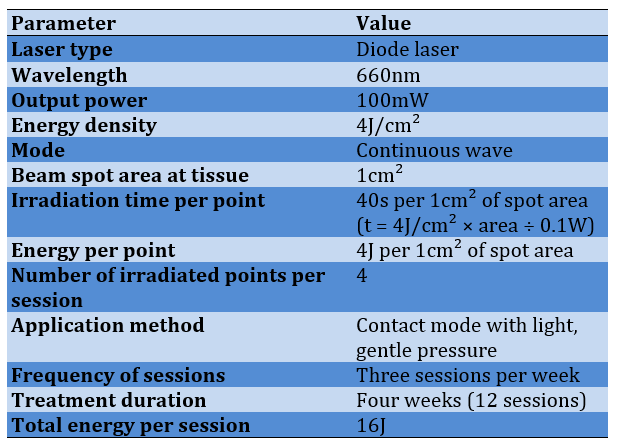
Group A additionally received LLLT with a diode laser system DLS-660 (MedLaser Tech.; USA) emitting at 660nm in continuous-wave mode at 100mW output and delivering an energy density of 4J/cm² per irradiated point. Irradiation was applied three times a week for four consecutive weeks (12 sessions), around the wound margins and over the wound bed (Table 1). This wavelength lies within the red band absorbed by cytochrome c oxidase [11], and the energy density falls within the 0.1–10J/cm² range reported as therapeutic in wound care [22]. In line with recommendations for reporting photomedicine dose [30], Table 1 lists the beam and dose parameters. Irradiation time per point follows from the relation
t = (energy density × spot area) / power
For a 1-cm² spot at 100mW and 4J/cm², this equals 40s and 4J per point (Contact mode with light gentle pressure; Output power was verified prior to each session using an external optical power meter). The operator and the patient wore protective eyewear during every session.
Table 1. Laser irradiation parameters used in the LLLT group (Both the operator and the patient wore protective eyewear during laser application. Parameters are reported according to the dose and beam-parameter checklist of Jenkins and Carroll)
Schedule of assessments
Participants attended baseline (week 0) and weekly visits at weeks 1, 2, 3, and 4. Wound photographs and clinical inflammation scores were recorded at every visit, whereas VAS pain, peri-wound temperature, and perfusion were recorded at baseline and week 4 and at each weekly visit, if applicable. Blood samples for TNF-α, IL-6, and CRP were collected only at baseline and week 4, and satisfaction and complete healing were assessed at week 4. Laser sessions in Group A took place three times a week, so that participants in this group also had contact with the treating team between assessment visits. Exploratory subgroup analyses were planned by wound duration (up to 14 days vs. longer) and by baseline wound area (below vs. above the median).
Clinical assessment: Wounds were evaluated at baseline and after the first, second, third, and fourth weeks of treatment. At each visit, the wound was photographed under standardized conditions (fixed camera-to-wound distance, uniform lighting, lens axis perpendicular to the wound plane, and an adjacent calibrated scale). Wound surface area was calculated with wound-measuring software; digital planimetry avoids the overestimation that occurs with the simple length-by-width method [31]. The percentage of wound healing was calculated as follows:
Wound healing (%) = [(initial wound area − follow-up wound area) / initial wound area] × 100
Complete healing was defined as full epithelialization without exudate. Clinical signs of inflammation (redness, swelling, exudation, and local pain) were recorded at each visit and graded on a four-point scale, from 0 (absent) to 3 (severe).
Biophysical assessment: The skin around the wound was assessed non-invasively. Peri-wound skin temperature was measured with an infrared thermometer, and local perfusion with laser Doppler flowmetry, an established method for assessing skin microcirculation [32]. Measurements were taken at standardized sites (e.g., 1cm from the wound margin) at baseline and week 4 in a climate-controlled room (25°C) after 15 minutes of acclimatization to limit environmental effects on skin blood flow.
Inflammatory biomarker assessment: Venous blood samples were collected from all participants at baseline and after four weeks of intervention. Serum was separated (3000×g for 10 minutes at 4°C) and stored at −80°C until analysis. TNF-α (Catalog numbers: DTA00D) and IL-6 (Catalog numbers: D6050) were measured by enzyme-linked immunosorbent assay (ELISA) with commercial kits (R&D Systems; Minneapolis, USA), following the manufacturer’s protocol. CRP was determined by a standard clinical laboratory procedure.
Pain and patient satisfaction
Pain intensity was rated by the patient on a 10-cm Visual Analog Scale (VAS; 0=no pain, 10=worst imaginable pain) [33] at baseline and week 4 and at each weekly visit, if applicable. Satisfaction with treatment was measured at the end of the treatment period using a structured scale, where higher scores indicate greater satisfaction with treatment effectiveness.
Ethical considerations
The study protocol was approved by the Institutional Ethics Committee of the Engineering Technical College of Najaf, Al-Furat Al-Awsat Technical University (Approval Code: ATU-ENG-2025-419) and conducted in accordance with the Declaration of Helsinki [34]. Written informed consent was obtained from all participants before enrollment. Patient confidentiality was maintained throughout the study, and participants were free to withdraw at any stage without any effect on their medical care.
Statistical analysis
Data were analyzed with SPSS 21 software. Normality was checked with the Shapiro–Wilk test and graphical inspection, and skewed cytokine concentrations were log-transformed. Continuous variables are presented as mean ± standard deviation (or median and interquartile range when non-normal) and categorical variables as frequencies and percentages. Baseline characteristics were compared with the independent-samples t-test (Mann–Whitney U test when non-normal) and the chi-square test (Fisher’s exact test when expected counts were below 5). For the primary outcome, percentage wound-area reduction at week 4 was compared between groups with the independent-samples t-test and confirmed by analysis of covariance adjusted for baseline wound area and wound duration. Healing over the four weeks was analyzed with repeated-measures analysis of variance or a linear mixed model testing the group × time interaction, with Bonferroni-adjusted pairwise comparisons. Within-group changes in biomarkers, pain, perfusion, and temperature were tested with a paired t-test, and between-group differences in change scores were tested with an independent-samples t-test or analysis of covariance adjusted for baseline value. Complete-healing rates were compared with the chi-square test. The association between percentage wound healing and changes in inflammatory markers was examined with Pearson’s correlation coefficient.
Findings
Participant flow and baseline characteristics
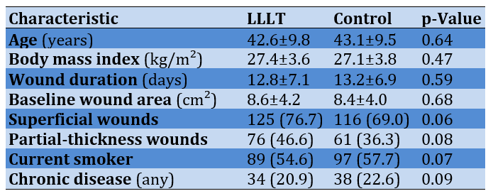
Three hundred and sixty women were randomized, 180 to each group. 17 participants in the LLLT group and 12 in the control group were removed due to clinical emergencies. All participants were women. As shown in Table 2, the two groups were not comparable at baseline in age, body mass index, wound duration, and wound area (p>0.05).
Table 2. Comparing the baseline demographic and clinical characteristics between the LLLT (n=163) and control (n=168) groups by Independent-samples t-test (Mann–Whitney U test if non-normal) or Chi-square test (Fisher’s exact test if expected counts <5)
Wound healing
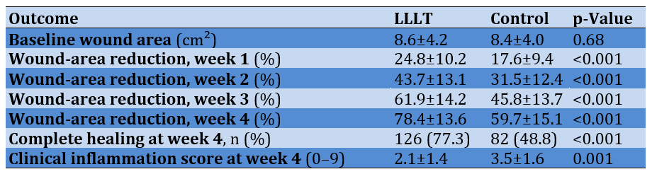
Table 3 shows the percentage wound-area reduction over the four-week follow-up. At week 4, the mean reduction was 78.4±13.6% (LLLT group) vs. 59.7±15.1% (control group), with a mean difference of 18.7% (95%CI: 15.8–21.6%; p<0.001; Cohen’s d=1.31). The repeated-measures analysis showed a significant group×time interaction (p=0.001), and between-group differences first became significant at week one.
Complete healing at week 4 was achieved by 126 (77.3%) participants in the LLLT group and 82 (48.8%) in the control group (risk ratio 1.54; 95%CI 1.27-1.86; p<0.001). Clinical inflammation scores (redness, swelling, exudation) were lower at week 4 in the LLLT group (p=0.001).
Table 3. Comparison of wound healing outcomes during the four-week follow-up in the LLLT (n=163) and control (n=168) groups by Independent-samples t-test for baseline wound area and wound duration and Chi-square test for complete healing
Inflammatory biomarkers
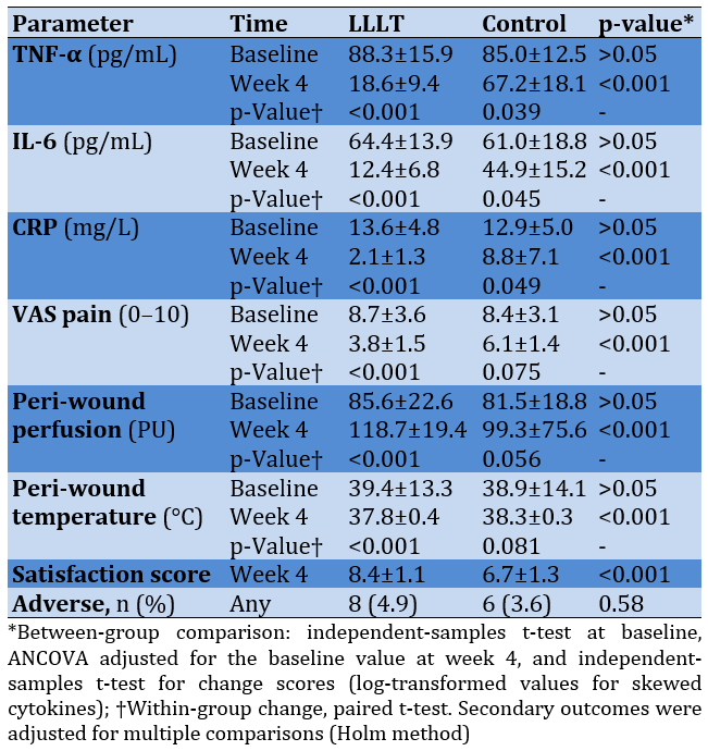
Serum concentrations at baseline and week 4 are presented in Table 4. Baseline TNF-α, IL-6, and CRP did not differ between groups (p>0.05). After four weeks, TNF-α changed by 69.7pg/mL in the LLLT group and by 17.8pg/mL in the control group (between-group p<0.001); the corresponding changes were 52.0 and 16.1pg/mL for IL-6 (p<0.001) and 11.5 and 4.1mg/L for CRP (p<0.001).
VAS pain fell from 8.7±3.6 to 3.8±1.5 in the LLLT group and from 8.4±3.1 to 6.1±1.4 in the control group (between-group p<0.001). Peri-wound perfusion changed by 33.1 perfusion units in the LLLT group and by 17.8 in the control group (p<0.001), and peri-wound temperature by -2.4°C and -0.6°C, respectively (p<0.001). The percentage of wound healing was negatively correlated with the change in TNF-α (r=-4.83; p=0.029), IL-6 (r=-6.08 p=0.008), and CRP (r=-3.68 p=0.045).
Table 4. Inflammatory biomarkers, pain, peri-wound perfusion and temperature, satisfaction, and adverse events at baseline and week 4
Exploratory subgroup analyses
Subgroup analyses revealed no significant differences in week-4 wound-area reduction stratified by baseline wound duration (up to 14 days vs. longer; interaction p=0.42) or baseline wound area (below vs. above the median; interaction p=0.31).
Discussion
This randomized controlled trial evaluated 660-nm LLLT as an adjunct to standard care in 360 women with superficial or partial-thickness skin wounds.
The primary outcome, wound closure, is best interpreted against a literature that has moved from skepticism to cautious support. An early review concluded that positive findings came mostly from small, poorly controlled series and that the evidence did not justify widespread use [19]. Since then, controlled human studies, including a triple-blind, sham-controlled trial in superficial wounds [20] and a randomized trial of light therapy in chronic diabetic wounds [21], have reported favorable healing outcomes, and a 2024 meta-analysis of 18 randomized trials found significantly greater wound-size reduction and healing rates with LLLT [25]. The present study contributes a large parallel-group sample, a fully specified irradiation protocol, and standardized planimetric measurement, addressing several methodological criticisms of earlier work. Healing proceeds through hemostasis, inflammation, proliferation, and remodeling, and disruption of any phase delays closure [2, 5]; an adjunct that acts on several phases at once is therefore biologically attractive.
The magnitude of the effect deserves comment. In the meta-analysis of randomized trials, the 95% confidence interval for the difference in percentage wound-size reduction between LLLT and control was 13.93 to 37.70 [25]. Because percentage area reduction depends on baseline size, and small wounds tend to close proportionally faster, adjusting for baseline wound area and wound duration, as we did in our analysis, is important when interpreting group differences.
The week-by-week trajectory is also informative. Changes in the first weeks mainly reflect the inflammatory phase and early granulation, whereas divergence between groups at weeks 3 and 4 would point to an effect on the proliferative phase, that is, fibroblast activity, angiogenesis, and re-epithelialization [2].
The biological rationale is consistent with the accepted mechanism of PBM. Red light is absorbed by cytochrome c oxidase, raising ATP synthesis and modulating nitric oxide, reactive oxygen species, and transcription factors [8-10]. The resulting increase in cellular energy supports fibroblast and keratinocyte proliferation and migration, collagen deposition, and neovascularization [12, 13, 23]. PBM also follows a biphasic dose–response pattern in which too little or too much light is ineffective [9], which is why complete dose reporting matters [30].
Inflammation was the second focus of the study. Persistent inflammatory signaling holds wounds in the inflammatory phase and prevents the shift to proliferation [3, 5], and TNF-α and IL-6 are prominent cytokines in this process [4]. PBM has been reported to lower these mediators [14]. A fall in TNF-α and IL-6 in the LLLT group would fit this pattern, and a parallel fall in CRP would be expected because IL-6 is the principal inducer of hepatic CRP synthesis [17]. CRP is widely used to monitor the inflammatory response [18], and combining it with cytokine measurements gives a fuller picture of the biological effect than clinical wound assessment alone. Interpretation should nevertheless be cautious: the wounds studied were small and localized, so systemic markers may change little and may also reflect wound-independent factors such as intercurrent infection or chronic disease. Targeting inflammatory, NF-κB, and oxidative pathways has been used to limit tissue injury in other conditions [15, 16], and a comparable principle may apply to cutaneous wounds.
Pain relief is an important secondary effect. PBM has been reported to relieve pain through several mechanisms, including modulating inflammatory mediators, improving tissue microcirculation, reducing edema, and altering peripheral nerve conduction [35]. Less pain can improve mobility, adherence to dressing changes and follow-up visits, and quality of life. The VAS is a simple, widely used patient-reported instrument [33], although its interpretation requires attention to concomitant analgesic use and expectation effects, because patients knew their allocation.
Tissue perfusion is a plausible mediator of the effect. Nitric oxide release after photon absorption can promote vasodilation and angiogenesis [10], and laser Doppler flowmetry offers a non-invasive window on skin microcirculation [32]. Better perfusion delivers oxygen and nutrients and removes metabolic waste, thereby supporting fibroblast function and granulation. Peri-wound temperature is an indirect indicator of both perfusion and inflammation and should be interpreted alongside the flux values.
The parameters used here (660nm, 100mW, continuous wave, 4J/cm²) fall within the range commonly reported, where red to near-infrared wavelengths and doses of roughly 0.1–10J/cm² have shown benefit [22]. Even so, differences in wavelength, power, dose, mode, and application technique are a major reason for inconsistent results between trials, and laser and LED devices are not interchangeable [26]. Power, energy, and treatment duration are interdependent design variables. Earlier work by the first author examined the relation between laser power and recovery period in endovenous laser ablation [36]; that application uses far higher energies and a different target tissue, but it illustrates why dose should be specified in reproducible terms and related to recovery time. Standardized reporting of all beam and dose parameters [30] is essential for comparison and meta-analysis. The relative merits of laser and LED sources have been reviewed [26]; LEDs cover larger areas at lower cost, whereas lasers offer collimated, narrow-band delivery, and the present findings apply only to laser delivery.
PBM and regenerative strategies share the aim of strengthening the body’s own repair mechanisms, and clinical improvement has been reported after stem-cell-based therapy in other conditions [27]. Combining PBM with cell-based products, platelet-rich plasma, growth factors, or bioengineered dressings is a logical next step, but it first requires quantifying the effect of PBM alone under controlled conditions, as attempted here.
From a public health perspective, delayed wound healing is costly and disabling [1], and evidence on simple, portable, low-risk adjuncts is directly relevant to equitable access to care. Because LLLT is non-invasive, requires little training once protocols are standardized, and can be delivered in outpatient settings, it may be a practical adjunct where specialized wound services are limited, including for populations affected by conflict-related injuries. The present trial did not enroll such patients, however, and its results should not be extrapolated to them without further study.
Safety is a further consideration. LLLT delivers non-thermogenic light [25], and no thermal injury is expected at the power and dose used here. Excluding participants with photosensitivity disorders, malignant lesions, and immunosuppressive treatment, along with mandatory eye protection during irradiation, reflects sensible precautions that should accompany clinical implementation.
Patient-centered outcomes complement the biological endpoints. Faster closure, less pain, and fewer dressing changes translate into fewer clinic visits, less infection-related anxiety, and an earlier return to daily activities, all of which contribute to satisfaction with care and quality of life. Satisfaction was measured here only at the end of treatment, and future trials should add validated quality-of-life instruments and health-economic endpoints to capture these benefits.
The study has several strengths: a prospective randomized design with a control group; a large sample; simultaneous assessment of clinical, biophysical, and laboratory inflammatory outcomes; a standardized and fully reported irradiation protocol; repeated weekly measurements throughout the treatment period; and a comprehensive outcome set that also captured pain, satisfaction, and adverse events.
Several limitations must also be acknowledged. First, the trial was conducted at a single center and follow-up lasted only four weeks, which precludes assessment of scar quality and recurrence. Second, only one wavelength and one irradiation mode were tested, so the findings cannot be generalized to other PBM systems. Third, the control group received no sham irradiation; participants could not be blinded, and placebo or attention effects on subjective outcomes such as pain and satisfaction cannot be excluded. Fourth, no histopathological or molecular analysis was performed, and growth factors, oxidative-stress markers, and collagen markers were not measured, so the mechanistic interpretation is inferential. Fifth, although randomization balances measured and unmeasured factors on average, differences in wound etiology, nutrition, smoking, chronic disease, and treatment adherence may still have influenced individual healing. Sixth, only women with superficial or partial-thickness wounds were enrolled, and deep wounds or severe tissue injury were excluded, which limits generalizability to men and to more complex wounds. Finally, because several secondary outcomes were analyzed, those results should be regarded as supportive rather than confirmatory.
For clinical practice, the results support using LLLT as a low-risk adjunct to standard care for superficial and partial-thickness wounds, provided protocols are standardized for wavelength, power, energy density, session duration, and frequency. Future work should include multicenter, sham-controlled, double-blind trials; head-to-head comparison of red and near-infrared wavelengths and of energy densities; long-term follow-up of scar quality and recurrence; histopathological and molecular studies of collagen synthesis, angiogenesis, fibroblast activity, cytokines, oxidative stress, and growth factors; trials in chronic diabetic, pressure, and venous ulcers and in burns; cost-effectiveness analyses; artificial-intelligence models to predict healing trajectories; and personalized PBM regimens matched to wound characteristics.
Conclusion
Adjunctive 660nm LLLT (100mW, 4J/cm², three sessions weekly for four weeks) improves wound closure at week 4 compared with standard care alone.
Acknowledgments: We thank all participants of this study, particularly the nurses, wound care center staff, laboratory workers, and laser therapy specialists who contributed directly or indirectly to this research.
Ethical Permissions: The research protocol was approved by the Research Ethics Committee of Research Ethics Committee of the Engineering Technical College of Najaf, Al-Furat Al-Awsat Technical University, Najaf, Iraq, (Approval Code: ATU-ENG-2025-419). The research was conducted in accordance with the ethical principles of the Declaration of Helsinki and its later amendments.
Conflicts of Interest: The authors declare no competing interests regarding the publication of this work. Artificial intelligence tools were used only for language editing. The authors are responsible for all scientific content, study design, data interpretation, analysis, and conclusions.
Authors’ Contribution: Yousif BG (First Author), Methodologist/Main Researcher/Discussion Writer (60%); Jassim SA (Second Author), Introduction Writer/Assistant Researcher/Statistical Analyst (40%)
Funding/Support: The authors declare that this research received no specific financial support from any government, commercial, or non-profit funding agencies.
Skin wounds are common, and impaired healing places a considerable burden on patients, health systems, and society. Delayed or incomplete healing prolongs hospitalization, increases the risk of recurrent infection, raises healthcare costs, and reduces the quality of life for affected individuals [1, 2]. Despite advances in wound care, restoring tissue integrity remains difficult, particularly in chronic wounds, where sustained inflammation and insufficient blood supply keep the tissue in a non-healing state [2, 3].
Normal healing is a highly orchestrated process that passes through four overlapping phases: hemostasis, inflammation, proliferation, and tissue remodeling [2]. Progression through these phases depends on efficient cellular communication, angiogenesis, extracellular matrix activity, collagen synthesis, and tight regulation of inflammatory mediators, including cytokines and growth factors [4, 5]. When one of these mechanisms fails, and especially when inflammation does not resolve in time, healing is prolonged, and the wound may become chronic [3, 5]. Standard wound care (cleansing, removal of devitalized tissue, and appropriate dressings) favors repair but does not directly stimulate the biology of healing, which explains the continuing interest in safe, non-invasive adjunctive therapies.
Low-level laser therapy (LLLT), now more commonly termed photobiomodulation (PBM), uses red or near-infrared light at intensities too low to cause thermal injury in order to modulate cellular function [6, 7]. The best-characterized mechanism is photon absorption by cytochrome c oxidase in the mitochondrial respiratory chain, which increases electron transport and adenosine triphosphate (ATP) production, releases nitric oxide, and alters reactive oxygen species signaling and transcription-factor activity [8-10]. The action spectra of cellular responses relevant to phototherapy show maxima in the red and near-infrared regions, consistent with absorption by this chromophore [11]. Downstream effects include fibroblast proliferation, collagen synthesis, keratinocyte migration, and angiogenesis, all central to tissue repair [12, 13].
PBM also influences inflammation. Experimental and clinical reports describe lower levels of pro-inflammatory mediators such as tumor necrosis factor-alpha (TNF-α) and interleukin-6 (IL-6), together with improved tissue oxygenation and microcirculation [14]. These effects are relevant because inflammation and oxidative stress are major mechanisms of tissue injury and delayed healing. In ischemic myocardial injury, suppressing the inflammatory response and apoptosis limits tissue damage [15], and in atherosclerosis, nuclear factor kappa-B (NF-κB) and oxidative pathways are therapeutic targets to reduce chronic inflammation [16]. C-reactive protein (CRP) is an acute-phase protein whose hepatic synthesis is driven mainly by IL-6, and it is widely used as a systemic marker of inflammatory activity [17]; raised levels have been reported in inflammatory conditions such as preeclampsia [18]. CRP was therefore included, together with TNF-α and IL-6, as a marker of the systemic inflammatory response.
The clinical evidence for LLLT in wound healing has nevertheless been uneven. An early critical review concluded that benefits seen in small case series had not been replicated in larger studies and that well-controlled trials with rational selection of laser parameters were needed [19]. Later work has been more encouraging. A triple-blind, sham-controlled study reported greater contraction of superficial wounds after LLLT [20], a randomized double-blind trial of LED phototherapy in chronic wounds of patients with diabetes reported promising results [21], and several reviews describe benefits in a broad range of wound types across a wide window of doses and wavelengths [22-24]. A recent meta-analysis of 18 randomized trials (670 wounds) found greater percentage reduction in wound size and higher healing rates with LLLT than without it [25]. Even so, studies differ widely in light source (laser or LED), wavelength, dose, and outcome definitions [26], and few combine clinical, biophysical, and biochemical endpoints in the same patients.
Regenerative approaches, including cell-based therapies, have likewise been reported to improve recovery by enhancing tissue regeneration [27]. Although these modalities differ from photobiomodulation, they share the aim of strengthening the body’s own repair capacity, and PBM is attractive because it is non-invasive and simple to deliver in outpatient settings.
A comprehensive evaluation is needed because wound area is a slow and indirect indicator of the processes that PBM is proposed to modify. Perfusion and cytokine measurements can show whether the anticipated mechanisms are engaged, while pain and satisfaction reflect benefit that matters to patients.
This randomized controlled trial was therefore designed to evaluate the effect of adjunctive 660-nm LLLT on skin wound healing, using standardized planimetric assessment of wound closure together with peri-wound perfusion, pain, and circulating inflammatory markers (TNF-α, IL-6, and CRP). We hypothesized that, compared with standard care alone, standard care plus LLLT would produce a greater reduction in wound area at week 4 and greater improvement in the secondary outcomes, without additional adverse effects.
Materials and Methods
Design and sampling
This prospective, two-arm, parallel-group study used a 1:1 allocation ratio and combined clinical, biophysical, and laboratory assessments. The report follows the CONSORT 2010 statement [28]. The study was conducted in the Outpatient Wound Care and Dermatology Units, Specialized Center, Najaf, Iraq. Recruitment and follow-up occurred over six months, from November 1, 2025, to May 1, 2026. All clinical observations, laser sessions, wound measurements, and laboratory tests followed a single written protocol. The sample size was calculated for the primary outcome, the difference in percentage wound-area reduction at week 4 between two independent groups, with a two-sided α of 0.05. A total of 175 participants per group provides 80% power to detect a standardized difference (Cohen’s d) of 0.3 [29]. Enrolling 180 participants per group (n=360) therefore gave adequate power for the primary comparison and about 90% power for d=0.35. Even with 10% loss to follow-up (162 analyzable participants per group), 80% power would be retained for d≈0.31.
Three hundred and sixty 18–65-year-old women with at least one superficial or partial-thickness skin wound that occurred in the last 1 to 30 days were enrolled. Wounds must be suitable for repeated standardized photography, and patients must be able to attend scheduled visits. Patients with uncontrolled diabetes mellitus, severe infection, malignant skin lesions, autoimmune disease, photosensitivity disorders, pregnancy or breastfeeding, previous laser treatment at the wound site, use of immunosuppressive medication, and deep wounds requiring surgical treatment were not selected. The samples were divided into two equal groups. Group A (LLLT group, n=180) received LLLT plus standard wound care, and Group B (control group, n=180) received standard wound care only. Participants were randomly allocated 1:1 using a computer-generated sequence prepared before enrollment by a researcher not involved in recruitment, treatment, or assessment. Allocation was concealed using sequentially numbered, opaque, sealed envelopes until baseline assessment was completed, to prevent selection bias. Because the control group did not receive sham irradiation, participants and laser operators could not be blinded. Wound photographs were coded and analyzed, and laboratory assays were run, by personnel unaware of group allocation.
Tools and Procedures
Standard wound care
Both groups received standard wound care, consisting of cleansing with sterile normal saline, removal of necrotic tissue, and application of sterile dressings. Topical growth factors and other advanced healing treatments were not used during the study period to avoid interference with the treatment effect. Concomitant medications, including analgesics, were recorded at each visit.
Group A additionally received LLLT with a diode laser system DLS-660 (MedLaser Tech.; USA) emitting at 660nm in continuous-wave mode at 100mW output and delivering an energy density of 4J/cm² per irradiated point. Irradiation was applied three times a week for four consecutive weeks (12 sessions), around the wound margins and over the wound bed (Table 1). This wavelength lies within the red band absorbed by cytochrome c oxidase [11], and the energy density falls within the 0.1–10J/cm² range reported as therapeutic in wound care [22]. In line with recommendations for reporting photomedicine dose [30], Table 1 lists the beam and dose parameters. Irradiation time per point follows from the relation
t = (energy density × spot area) / power
For a 1-cm² spot at 100mW and 4J/cm², this equals 40s and 4J per point (Contact mode with light gentle pressure; Output power was verified prior to each session using an external optical power meter). The operator and the patient wore protective eyewear during every session.
Table 1. Laser irradiation parameters used in the LLLT group (Both the operator and the patient wore protective eyewear during laser application. Parameters are reported according to the dose and beam-parameter checklist of Jenkins and Carroll)
Schedule of assessments
Participants attended baseline (week 0) and weekly visits at weeks 1, 2, 3, and 4. Wound photographs and clinical inflammation scores were recorded at every visit, whereas VAS pain, peri-wound temperature, and perfusion were recorded at baseline and week 4 and at each weekly visit, if applicable. Blood samples for TNF-α, IL-6, and CRP were collected only at baseline and week 4, and satisfaction and complete healing were assessed at week 4. Laser sessions in Group A took place three times a week, so that participants in this group also had contact with the treating team between assessment visits. Exploratory subgroup analyses were planned by wound duration (up to 14 days vs. longer) and by baseline wound area (below vs. above the median).
Clinical assessment: Wounds were evaluated at baseline and after the first, second, third, and fourth weeks of treatment. At each visit, the wound was photographed under standardized conditions (fixed camera-to-wound distance, uniform lighting, lens axis perpendicular to the wound plane, and an adjacent calibrated scale). Wound surface area was calculated with wound-measuring software; digital planimetry avoids the overestimation that occurs with the simple length-by-width method [31]. The percentage of wound healing was calculated as follows:
Wound healing (%) = [(initial wound area − follow-up wound area) / initial wound area] × 100
Complete healing was defined as full epithelialization without exudate. Clinical signs of inflammation (redness, swelling, exudation, and local pain) were recorded at each visit and graded on a four-point scale, from 0 (absent) to 3 (severe).
Biophysical assessment: The skin around the wound was assessed non-invasively. Peri-wound skin temperature was measured with an infrared thermometer, and local perfusion with laser Doppler flowmetry, an established method for assessing skin microcirculation [32]. Measurements were taken at standardized sites (e.g., 1cm from the wound margin) at baseline and week 4 in a climate-controlled room (25°C) after 15 minutes of acclimatization to limit environmental effects on skin blood flow.
Inflammatory biomarker assessment: Venous blood samples were collected from all participants at baseline and after four weeks of intervention. Serum was separated (3000×g for 10 minutes at 4°C) and stored at −80°C until analysis. TNF-α (Catalog numbers: DTA00D) and IL-6 (Catalog numbers: D6050) were measured by enzyme-linked immunosorbent assay (ELISA) with commercial kits (R&D Systems; Minneapolis, USA), following the manufacturer’s protocol. CRP was determined by a standard clinical laboratory procedure.
Pain and patient satisfaction
Pain intensity was rated by the patient on a 10-cm Visual Analog Scale (VAS; 0=no pain, 10=worst imaginable pain) [33] at baseline and week 4 and at each weekly visit, if applicable. Satisfaction with treatment was measured at the end of the treatment period using a structured scale, where higher scores indicate greater satisfaction with treatment effectiveness.
Ethical considerations
The study protocol was approved by the Institutional Ethics Committee of the Engineering Technical College of Najaf, Al-Furat Al-Awsat Technical University (Approval Code: ATU-ENG-2025-419) and conducted in accordance with the Declaration of Helsinki [34]. Written informed consent was obtained from all participants before enrollment. Patient confidentiality was maintained throughout the study, and participants were free to withdraw at any stage without any effect on their medical care.
Statistical analysis
Data were analyzed with SPSS 21 software. Normality was checked with the Shapiro–Wilk test and graphical inspection, and skewed cytokine concentrations were log-transformed. Continuous variables are presented as mean ± standard deviation (or median and interquartile range when non-normal) and categorical variables as frequencies and percentages. Baseline characteristics were compared with the independent-samples t-test (Mann–Whitney U test when non-normal) and the chi-square test (Fisher’s exact test when expected counts were below 5). For the primary outcome, percentage wound-area reduction at week 4 was compared between groups with the independent-samples t-test and confirmed by analysis of covariance adjusted for baseline wound area and wound duration. Healing over the four weeks was analyzed with repeated-measures analysis of variance or a linear mixed model testing the group × time interaction, with Bonferroni-adjusted pairwise comparisons. Within-group changes in biomarkers, pain, perfusion, and temperature were tested with a paired t-test, and between-group differences in change scores were tested with an independent-samples t-test or analysis of covariance adjusted for baseline value. Complete-healing rates were compared with the chi-square test. The association between percentage wound healing and changes in inflammatory markers was examined with Pearson’s correlation coefficient.
Findings
Participant flow and baseline characteristics
Three hundred and sixty women were randomized, 180 to each group. 17 participants in the LLLT group and 12 in the control group were removed due to clinical emergencies. All participants were women. As shown in Table 2, the two groups were not comparable at baseline in age, body mass index, wound duration, and wound area (p>0.05).
Table 2. Comparing the baseline demographic and clinical characteristics between the LLLT (n=163) and control (n=168) groups by Independent-samples t-test (Mann–Whitney U test if non-normal) or Chi-square test (Fisher’s exact test if expected counts <5)
Wound healing
Table 3 shows the percentage wound-area reduction over the four-week follow-up. At week 4, the mean reduction was 78.4±13.6% (LLLT group) vs. 59.7±15.1% (control group), with a mean difference of 18.7% (95%CI: 15.8–21.6%; p<0.001; Cohen’s d=1.31). The repeated-measures analysis showed a significant group×time interaction (p=0.001), and between-group differences first became significant at week one.
Complete healing at week 4 was achieved by 126 (77.3%) participants in the LLLT group and 82 (48.8%) in the control group (risk ratio 1.54; 95%CI 1.27-1.86; p<0.001). Clinical inflammation scores (redness, swelling, exudation) were lower at week 4 in the LLLT group (p=0.001).
Table 3. Comparison of wound healing outcomes during the four-week follow-up in the LLLT (n=163) and control (n=168) groups by Independent-samples t-test for baseline wound area and wound duration and Chi-square test for complete healing
Inflammatory biomarkers
Serum concentrations at baseline and week 4 are presented in Table 4. Baseline TNF-α, IL-6, and CRP did not differ between groups (p>0.05). After four weeks, TNF-α changed by 69.7pg/mL in the LLLT group and by 17.8pg/mL in the control group (between-group p<0.001); the corresponding changes were 52.0 and 16.1pg/mL for IL-6 (p<0.001) and 11.5 and 4.1mg/L for CRP (p<0.001).
VAS pain fell from 8.7±3.6 to 3.8±1.5 in the LLLT group and from 8.4±3.1 to 6.1±1.4 in the control group (between-group p<0.001). Peri-wound perfusion changed by 33.1 perfusion units in the LLLT group and by 17.8 in the control group (p<0.001), and peri-wound temperature by -2.4°C and -0.6°C, respectively (p<0.001). The percentage of wound healing was negatively correlated with the change in TNF-α (r=-4.83; p=0.029), IL-6 (r=-6.08 p=0.008), and CRP (r=-3.68 p=0.045).
Table 4. Inflammatory biomarkers, pain, peri-wound perfusion and temperature, satisfaction, and adverse events at baseline and week 4
Exploratory subgroup analyses
Subgroup analyses revealed no significant differences in week-4 wound-area reduction stratified by baseline wound duration (up to 14 days vs. longer; interaction p=0.42) or baseline wound area (below vs. above the median; interaction p=0.31).
Discussion
This randomized controlled trial evaluated 660-nm LLLT as an adjunct to standard care in 360 women with superficial or partial-thickness skin wounds.
The primary outcome, wound closure, is best interpreted against a literature that has moved from skepticism to cautious support. An early review concluded that positive findings came mostly from small, poorly controlled series and that the evidence did not justify widespread use [19]. Since then, controlled human studies, including a triple-blind, sham-controlled trial in superficial wounds [20] and a randomized trial of light therapy in chronic diabetic wounds [21], have reported favorable healing outcomes, and a 2024 meta-analysis of 18 randomized trials found significantly greater wound-size reduction and healing rates with LLLT [25]. The present study contributes a large parallel-group sample, a fully specified irradiation protocol, and standardized planimetric measurement, addressing several methodological criticisms of earlier work. Healing proceeds through hemostasis, inflammation, proliferation, and remodeling, and disruption of any phase delays closure [2, 5]; an adjunct that acts on several phases at once is therefore biologically attractive.
The magnitude of the effect deserves comment. In the meta-analysis of randomized trials, the 95% confidence interval for the difference in percentage wound-size reduction between LLLT and control was 13.93 to 37.70 [25]. Because percentage area reduction depends on baseline size, and small wounds tend to close proportionally faster, adjusting for baseline wound area and wound duration, as we did in our analysis, is important when interpreting group differences.
The week-by-week trajectory is also informative. Changes in the first weeks mainly reflect the inflammatory phase and early granulation, whereas divergence between groups at weeks 3 and 4 would point to an effect on the proliferative phase, that is, fibroblast activity, angiogenesis, and re-epithelialization [2].
The biological rationale is consistent with the accepted mechanism of PBM. Red light is absorbed by cytochrome c oxidase, raising ATP synthesis and modulating nitric oxide, reactive oxygen species, and transcription factors [8-10]. The resulting increase in cellular energy supports fibroblast and keratinocyte proliferation and migration, collagen deposition, and neovascularization [12, 13, 23]. PBM also follows a biphasic dose–response pattern in which too little or too much light is ineffective [9], which is why complete dose reporting matters [30].
Inflammation was the second focus of the study. Persistent inflammatory signaling holds wounds in the inflammatory phase and prevents the shift to proliferation [3, 5], and TNF-α and IL-6 are prominent cytokines in this process [4]. PBM has been reported to lower these mediators [14]. A fall in TNF-α and IL-6 in the LLLT group would fit this pattern, and a parallel fall in CRP would be expected because IL-6 is the principal inducer of hepatic CRP synthesis [17]. CRP is widely used to monitor the inflammatory response [18], and combining it with cytokine measurements gives a fuller picture of the biological effect than clinical wound assessment alone. Interpretation should nevertheless be cautious: the wounds studied were small and localized, so systemic markers may change little and may also reflect wound-independent factors such as intercurrent infection or chronic disease. Targeting inflammatory, NF-κB, and oxidative pathways has been used to limit tissue injury in other conditions [15, 16], and a comparable principle may apply to cutaneous wounds.
Pain relief is an important secondary effect. PBM has been reported to relieve pain through several mechanisms, including modulating inflammatory mediators, improving tissue microcirculation, reducing edema, and altering peripheral nerve conduction [35]. Less pain can improve mobility, adherence to dressing changes and follow-up visits, and quality of life. The VAS is a simple, widely used patient-reported instrument [33], although its interpretation requires attention to concomitant analgesic use and expectation effects, because patients knew their allocation.
Tissue perfusion is a plausible mediator of the effect. Nitric oxide release after photon absorption can promote vasodilation and angiogenesis [10], and laser Doppler flowmetry offers a non-invasive window on skin microcirculation [32]. Better perfusion delivers oxygen and nutrients and removes metabolic waste, thereby supporting fibroblast function and granulation. Peri-wound temperature is an indirect indicator of both perfusion and inflammation and should be interpreted alongside the flux values.
The parameters used here (660nm, 100mW, continuous wave, 4J/cm²) fall within the range commonly reported, where red to near-infrared wavelengths and doses of roughly 0.1–10J/cm² have shown benefit [22]. Even so, differences in wavelength, power, dose, mode, and application technique are a major reason for inconsistent results between trials, and laser and LED devices are not interchangeable [26]. Power, energy, and treatment duration are interdependent design variables. Earlier work by the first author examined the relation between laser power and recovery period in endovenous laser ablation [36]; that application uses far higher energies and a different target tissue, but it illustrates why dose should be specified in reproducible terms and related to recovery time. Standardized reporting of all beam and dose parameters [30] is essential for comparison and meta-analysis. The relative merits of laser and LED sources have been reviewed [26]; LEDs cover larger areas at lower cost, whereas lasers offer collimated, narrow-band delivery, and the present findings apply only to laser delivery.
PBM and regenerative strategies share the aim of strengthening the body’s own repair mechanisms, and clinical improvement has been reported after stem-cell-based therapy in other conditions [27]. Combining PBM with cell-based products, platelet-rich plasma, growth factors, or bioengineered dressings is a logical next step, but it first requires quantifying the effect of PBM alone under controlled conditions, as attempted here.
From a public health perspective, delayed wound healing is costly and disabling [1], and evidence on simple, portable, low-risk adjuncts is directly relevant to equitable access to care. Because LLLT is non-invasive, requires little training once protocols are standardized, and can be delivered in outpatient settings, it may be a practical adjunct where specialized wound services are limited, including for populations affected by conflict-related injuries. The present trial did not enroll such patients, however, and its results should not be extrapolated to them without further study.
Safety is a further consideration. LLLT delivers non-thermogenic light [25], and no thermal injury is expected at the power and dose used here. Excluding participants with photosensitivity disorders, malignant lesions, and immunosuppressive treatment, along with mandatory eye protection during irradiation, reflects sensible precautions that should accompany clinical implementation.
Patient-centered outcomes complement the biological endpoints. Faster closure, less pain, and fewer dressing changes translate into fewer clinic visits, less infection-related anxiety, and an earlier return to daily activities, all of which contribute to satisfaction with care and quality of life. Satisfaction was measured here only at the end of treatment, and future trials should add validated quality-of-life instruments and health-economic endpoints to capture these benefits.
The study has several strengths: a prospective randomized design with a control group; a large sample; simultaneous assessment of clinical, biophysical, and laboratory inflammatory outcomes; a standardized and fully reported irradiation protocol; repeated weekly measurements throughout the treatment period; and a comprehensive outcome set that also captured pain, satisfaction, and adverse events.
Several limitations must also be acknowledged. First, the trial was conducted at a single center and follow-up lasted only four weeks, which precludes assessment of scar quality and recurrence. Second, only one wavelength and one irradiation mode were tested, so the findings cannot be generalized to other PBM systems. Third, the control group received no sham irradiation; participants could not be blinded, and placebo or attention effects on subjective outcomes such as pain and satisfaction cannot be excluded. Fourth, no histopathological or molecular analysis was performed, and growth factors, oxidative-stress markers, and collagen markers were not measured, so the mechanistic interpretation is inferential. Fifth, although randomization balances measured and unmeasured factors on average, differences in wound etiology, nutrition, smoking, chronic disease, and treatment adherence may still have influenced individual healing. Sixth, only women with superficial or partial-thickness wounds were enrolled, and deep wounds or severe tissue injury were excluded, which limits generalizability to men and to more complex wounds. Finally, because several secondary outcomes were analyzed, those results should be regarded as supportive rather than confirmatory.
For clinical practice, the results support using LLLT as a low-risk adjunct to standard care for superficial and partial-thickness wounds, provided protocols are standardized for wavelength, power, energy density, session duration, and frequency. Future work should include multicenter, sham-controlled, double-blind trials; head-to-head comparison of red and near-infrared wavelengths and of energy densities; long-term follow-up of scar quality and recurrence; histopathological and molecular studies of collagen synthesis, angiogenesis, fibroblast activity, cytokines, oxidative stress, and growth factors; trials in chronic diabetic, pressure, and venous ulcers and in burns; cost-effectiveness analyses; artificial-intelligence models to predict healing trajectories; and personalized PBM regimens matched to wound characteristics.
Conclusion
Adjunctive 660nm LLLT (100mW, 4J/cm², three sessions weekly for four weeks) improves wound closure at week 4 compared with standard care alone.
Acknowledgments: We thank all participants of this study, particularly the nurses, wound care center staff, laboratory workers, and laser therapy specialists who contributed directly or indirectly to this research.
Ethical Permissions: The research protocol was approved by the Research Ethics Committee of Research Ethics Committee of the Engineering Technical College of Najaf, Al-Furat Al-Awsat Technical University, Najaf, Iraq, (Approval Code: ATU-ENG-2025-419). The research was conducted in accordance with the ethical principles of the Declaration of Helsinki and its later amendments.
Conflicts of Interest: The authors declare no competing interests regarding the publication of this work. Artificial intelligence tools were used only for language editing. The authors are responsible for all scientific content, study design, data interpretation, analysis, and conclusions.
Authors’ Contribution: Yousif BG (First Author), Methodologist/Main Researcher/Discussion Writer (60%); Jassim SA (Second Author), Introduction Writer/Assistant Researcher/Statistical Analyst (40%)
Funding/Support: The authors declare that this research received no specific financial support from any government, commercial, or non-profit funding agencies.
References
1. Sen CK. Human wounds and its burden: An updated compendium of estimates. Adv Wound Care. 2019;8(2):39-48. [Link] [DOI:10.1089/wound.2019.0946]
2. Rodrigues M, Kosaric N, Bonham CA, Gurtner GC. Wound healing: A cellular perspective. Physiol Rev. 2019;99(1):665-706. [Link] [DOI:10.1152/physrev.00067.2017]
3. Frykberg RG, Banks J. Challenges in the treatment of chronic wounds. Adv Wound Care. 2015;4(9):560-82. [Link] [DOI:10.1089/wound.2015.0635]
4. Barrientos S, Stojadinovic O, Golinko MS, Brem H, Tomic-Canic M. Growth factors and cytokines in wound healing. Wound Repair Regen. 2008;16(5):585-601. [Link] [DOI:10.1111/j.1524-475X.2008.00410.x]
5. Eming SA, Martin P, Tomic-Canic M. Wound repair and regeneration: Mechanisms, signaling, and translation. Sci Transl Med. 2014;6(265):265sr6. [Link] [DOI:10.1126/scitranslmed.3009337]
6. Hamblin MR. Photobiomodulation or low-level laser therapy. J Biophotonics. 2016;9(11-12):1122-4. [Link] [DOI:10.1002/jbio.201670113]
7. Anders JJ, Lanzafame RJ, Arany PR. Low-level light/laser therapy versus photobiomodulation therapy. Photomed Laser Surg. 2015;33(4):183-4. [Link] [DOI:10.1089/pho.2015.9848]
8. Karu T. Mitochondrial mechanisms of photobiomodulation in context of new data about multiple roles of ATP. Photomed Laser Surg. 2010;28(2):159-60. [Link] [DOI:10.1089/pho.2010.2789]
9. Chung H, Dai T, Sharma SK, Huang YY, Carroll JD, Hamblin MR. The nuts and bolts of low-level laser (light) therapy. Ann Biomed Eng. 2012;40(2):516-33. [Link] [DOI:10.1007/s10439-011-0454-7]
10. De Freitas LF, Hamblin MR. Proposed mechanisms of photobiomodulation or low-level light therapy. IEEE J Sel Top Quantum Electron. 2016;22(3):7000417. [Link] [DOI:10.1109/JSTQE.2016.2561201]
11. Karu TI, Kolyakov SF. Exact action spectra for cellular responses relevant to phototherapy. Photobiomodulation Photomed Laser Surg. 2005;23(4):355-61. [Link] [DOI:10.1089/pho.2005.23.355]
12. Avci P, Gupta A, Sadasivam M, Vecchio D, Pam Z, Pam N, et al. Low-level laser (light) therapy (LLLT) in skin: Stimulating, healing, restoring. Semin Cutan Med Surg. 2013;32(1):41-52. [Link]
13. Dompe C, Moncrieff L, Matys J, Grzech-Lesniak K, Kocherova I, Bryja A, et al. Photobiomodulation-Underlying mechanism and clinical applications. J Clin Med. 2020;9(6):1724. [Link] [DOI:10.3390/jcm9061724]
14. Hamblin MR. Mechanisms and applications of the anti-inflammatory effects of photobiomodulation. AIMS Biophys. 2017;4(3):337-61. [Link] [DOI:10.3934/biophy.2017.3.337]
15. Hadi NR, Al-Amran FGY, Yousif MG, Hussein FH. Amelioration of myocardial ischemia/reperfusion injury by L-carnitine via down-regulation of the inflammatory response and apoptosis. Exp Clin Cardiol. 2014;20(8):3732-40. [Link]
16. Hadi NR, Yousif NG, Abdulzahra MS, Mohammad BI, Al-Amran FG, Majeed ML, et al. Role of NF-kB and oxidative pathways in atherosclerosis: Cross-talk between dyslipidemia and candesartan. Cardiovasc Ther. 2013;31(6):381-7. [Link] [DOI:10.1111/1755-5922.12033]
17. Pepys MB, Hirschfield GM. C-reactive protein: A critical update. J Clin Invest. 2003;111(12):1805-12. [Link] [DOI:10.1172/JCI200318921]
18. Sadiq AM, Hussein CM, Yousif MG, Mohammed RJ. Correlation between highly sensitive C-reactive protein level in cases of preeclampsia with or without intrauterine-growth restriction. Indian J Forensic Med Toxicol. 2020;14(4):6012-6. [Link]
19. Posten W, Wrone DA, Dover JS, Arndt KA, Silapunt S, Alam M. Low-level laser therapy for wound healing: Mechanism and efficacy. Dermatol Surg. 2005;31(3):334-40. [Link] [DOI:10.1097/00042728-200503000-00016]
20. Hopkins JT, McLoda TA, Seegmiller JG, Baxter GD. Low-level laser therapy facilitates superficial wound healing in humans: A triple-blind, sham-controlled study. J Athl Train. 2004;39(3):223-9. [Link]
21. Frangez I, Nizic-Kos T, Frangez HB. Phototherapy with LED shows promising results in healing chronic wounds in diabetes mellitus patients: A prospective randomized double-blind study. Photomed Laser Surg. 2018;36(7):377-82. [Link] [DOI:10.1089/pho.2017.4382]
22. Mosca RC, Ong AA, Albasha O, Bass K, Arany P. Photobiomodulation therapy for wound care: A potent, noninvasive, photoceutical approach. Adv Skin Wound Care. 2019;32(4):157-67. [Link] [DOI:10.1097/01.ASW.0000553600.97572.d2]
23. Kuffler DP. Photobiomodulation in promoting wound healing: A review. Regen Med. 2016;11(1):107-22. [Link] [DOI:10.2217/rme.15.82]
24. Aggarwal I, Lio PA. Photobiomodulation therapy and low-level light therapy in wound healing. Lasers Med Sci. 2023;38(1):239. [Link] [DOI:10.1007/s10103-023-03909-9]
25. Taha N, Daoud H, Malik T, Shettysowkoor J, Rahman S. The effects of low-level laser therapy on wound healing and pain management in skin wounds: A systematic review and meta-analysis. Cureus. 2024;16(10):e72542. [Link] [DOI:10.7759/cureus.72542]
26. Chaves MEA, Araujo AR, Piancastelli ACC, Pinotti M. Effects of low-power light therapy on wound healing: LASER x LED. ANAIS BRASILEIROS DE DERMATOLOGIA. 2014;89(4):616-23. [Link] [DOI:10.1590/abd1806-4841.20142519]
27. Yousif NG, Yousif MG, Mohsen AAU, El-Bakaa HS, Younise MH, Altimimi AN, et al. Prospective single center analysis of outcome stem cells transplants in patients with cerebral palsy. Pol Merkur Lekarski. 2023;51(4):339-45. [Link] [DOI:10.36740/Merkur202304107]
28. Schulz KF, Altman DG, Moher D, CONSORT Group. CONSORT 2010 statement: Updated guidelines for reporting parallel group randomised trials. BMJ. 2010;340:c332. [Link] [DOI:10.1136/bmj.c332]
29. Cohen J. Statistical power analysis for the behavioral sciences. 2nd ed. Hillsdale (NJ): Lawrence Erlbaum Associates; 1988. [Link]
30. Jenkins PA, Carroll JD. How to report low-level laser therapy (LLLT)/photomedicine dose and beam parameters in clinical and laboratory studies. Photomed Laser Surg. 2011;29(12):785-7. [Link] [DOI:10.1089/pho.2011.9895]
31. Rogers LC, Bevilacqua NJ, Armstrong DG, Andros G. Digital planimetry results in more accurate wound measurements: A comparison to standard ruler measurements. J Diabetes Sci Technol. 2010;4(4):799-802. [Link] [DOI:10.1177/193229681000400405]
32. Roustit M, Cracowski JL. Assessment of endothelial and neurovascular function in human skin microcirculation. Trends Pharmacol Sci. 2013;34(7):373-84. [Link] [DOI:10.1016/j.tips.2013.05.007]
33. Hawker GA, Mian S, Kendzerska T, French M. Measures of adult pain: Visual Analog Scale for Pain (VAS Pain), Numeric Rating Scale for Pain (NRS Pain), McGill Pain Questionnaire (MPQ), Short-Form McGill Pain Questionnaire (SF-MPQ), Chronic Pain Grade Scale (CPGS), Short Form-36 Bodily Pain Scale (SF-36 BPS), and Measure of Intermittent and Constant Osteoarthritis Pain (ICOAP). Arthritis Care Res. 2011;63(Suppl 11):S240-52. [Link] [DOI:10.1002/acr.20543]
34. World Medical Association. World Medical Association Declaration of Helsinki: Ethical principles for medical research involving human subjects. JAMA. 2013;310(20):2191-4. [Link] [DOI:10.1001/jama.2013.281053]
35. Bjordal JM, Johnson MI, Iversen V, Aimbire F, Lopes-Martins RA. Low-level laser therapy in acute pain: A systematic review of possible mechanisms of action and clinical effects in randomized placebo-controlled trials. Photomed Laser Surg. 2006;24(2):158-68. [Link] [DOI:10.1089/pho.2006.24.158]
36. Yousif BG, Abdul-Wahid SN, Shabaa KJ. Predict the laser power and recovery period in treatment by endovenous laser ablation (EVLA). Baghdad Sci J. 2024;21(2):473-9. [Link] [DOI:10.21123/bsj.2023.7825]